 Login / Register


- Clone
- 11C12E12 (See other available formats)
- Regulatory Status
- RUO
- Other Names
- S-100 protein beta chain, S-100 protein subunit beta, S100 calcium-binding protein B
- Isotype
- Mouse IgG1
- Ave. Rating
- Submit a Review
- Product Citations
- publications

-

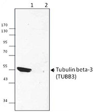
Western blot analysis of rat brain (lane 1) and mouse brain (lane 2). Anti- S100B antibody (clone 11C12E12 ) was used, followed by HRP Goat anti-mouse IgG (minimal x-reactivity) antibody. Purified anti-GAPDH antibody was used as a loading control. We are not sure about the specificity of the high molecular weight band. -

SeqIF™ (sequential immunofluorescence) staining on COMET™ of Purified anti-S100B (clone 11C12E12, yellow) on formalin-fixed paraffin-embedded human pancreatic carcinoma at 1.25 µg/mL. Alexa Fluor™ Plus 647 Goat anti-Mouse IgG antibody (Lunaphore, Cat. No. DR647MS) was used as a secondary antibody. Nuclei were counterstained with DAPI (blue). Tissue underwent an all-in-one dewaxing and antigen retrieval preprocessing.
| Cat # | Size | Price | Quantity Check Availability | Save | ||
|---|---|---|---|---|---|---|
| 676603 | 25 µg | 81€ | ||||
| 676604 | 100 µg | 203€ | ||||
S100B is glial-specific. It has been shown that it is only expressed by a subtype of mature astrocytes that ensheath blood vessels, and by NG2-expressing cells. This protein may function in neurite extension, stimulation of Ca2+ fluxes, inhibition of PKC-mediated phosphorylation, astrocytosis and axonal proliferation, and inhibition of microtubule assembly. In the developing central nervous system (CNS), it acts as a neurotrophic factor and neuronal survival protein. In the adult organism, it is usually elevated due to nervous system damage, which makes it a potential clinical marker. S100B is secreted by astrocytes or can spill from injured cells and enter the extracellular space or bloodstream. Serum levels of S100B increase in patients during the acute phase of brain damage. Over the last decade, S100B has emerged as a candidate peripheral biomarker of blood–brain barrier (BBB) permeability and central nervous system injury.
Product DetailsProduct Details
- Verified Reactivity
- Human, Mouse, Rat
- Antibody Type
- Monoclonal
- Host Species
- Mouse
- Immunogen
- Full length recombinant human S100B protein.
- Formulation
- Phosphate-buffered solution, pH 7.2, containing 0.09% sodium azide.
- Preparation
- The antibody was purified by affinity chromatography.
- Concentration
- 0.5 mg/ml
- Storage & Handling
- The antibody solution should be stored undiluted between 2°C and 8°C.
- Application
-
WB - Quality tested
SB - Community verified - Recommended Usage
-
Each lot of this antibody is quality control tested by Western blotting. For Western blotting, the suggested use of this reagent is 0.5 - 2.0 µg per ml. It is recommended that the reagent be titrated for optimal performance for each application.
- Additional Product Notes
-
For the use of this antibody in spatial biology applications, we have partnered with Lunaphore Technologies for demonstration of our antibodies on the COMET™. The COMET™ platform is an automated, end-to-end spatial biology solution developed for rapid and flexible multiplex tissue profiling. More information on the COMET™ and a complete list of our antibodies that have been demonstrated on the COMET™ can be found here.
- Product Citations
-
- RRID
-
AB_2734531 (BioLegend Cat. No. 676603)
AB_2571890 (BioLegend Cat. No. 676604)
Antigen Details
- Structure
- 92 amino acids with a predicted molecular weight of approximately 10.7 kD.
- Distribution
-
Mainly expressed in mature astrocytes of the brain. It is also found in a variety of other cell types such as T lymphocytes, monocytes, and platelets.
- Function
- Neurite extension, stimulation of Ca2+ fluxes, inhibition of PKC-mediated phosphorylation, astrocytosis and axonal proliferation, and inhibition of microtubule assembly.
- Interaction
- STK38, CACYBP, ATAD3A, S100A6, CAPZA1 AGER, and PPP5C.
- Cell Type
- Astrocytes
- Biology Area
- Cell Biology, Immunology, Neuroinflammation, Neuroscience, Neuroscience Cell Markers, Signal Transduction
- Antigen References
-
1. Wang D, et al. 2008. Prog. Neurobiol. 86:342.
2. Michetti F, et al. 2012. J. Neurochem. 120:644.
3. Zongo D, et al. 2012. Annals. of Emergency Medicine 59:209.
4. Deloulme J, et al. 2000. J. Biol. Chem. 275:35302-10. - Gene ID
- 6285 View all products for this Gene ID
- UniProt
- View information about S100B on UniProt.org

Related FAQs
- If an antibody clone has been previously successfully used in IBEX in one fluorescent format, will other antibody formats work as well?
-
It’s likely that other fluorophore conjugates to the same antibody clone will also be compatible with IBEX using the same sample fixation procedure. Ultimately a directly conjugated antibody’s utility in fluorescent imaging and IBEX may be specific to the sample and microscope being used in the experiment. Some antibody clone conjugates may perform better than others due to performance differences in non-specific binding, fluorophore brightness, and other biochemical properties unique to that conjugate.
- Will antibodies my lab is already using for fluorescent or chromogenic IHC work in IBEX?
-
Fundamentally, IBEX as a technique that works much in the same way as single antibody panels or single marker IF/IHC. If you’re already successfully using an antibody clone on a sample of interest, it is likely that clone will have utility in IBEX. It is expected some optimization and testing of different antibody fluorophore conjugates will be required to find a suitable format; however, legacy microscopy techniques like chromogenic IHC on fixed or frozen tissue is an excellent place to start looking for useful antibodies.
- Are other fluorophores compatible with IBEX?
-
Over 18 fluorescent formats have been screened for use in IBEX, however, it is likely that other fluorophores are able to be rapidly bleached in IBEX. If a fluorophore format is already suitable for your imaging platform it can be tested for compatibility in IBEX.
- The same antibody works in one tissue type but not another. What is happening?
-
Differences in tissue properties may impact both the ability of an antibody to bind its target specifically and impact the ability of a specific fluorophore conjugate to overcome the background fluorescent signal in a given tissue. Secondary stains, as well as testing multiple fluorescent conjugates of the same clone, may help to troubleshoot challenging targets or tissues. Using a reference control tissue may also give confidence in the specificity of your staining.
- How can I be sure the staining I’m seeing in my tissue is real?
-
In general, best practices for validating an antibody in traditional chromogenic or fluorescent IHC are applicable to IBEX. Please reference the Nature Methods review on antibody based multiplexed imaging for resources on validating antibodies for IBEX.
Other Formats
View All S100B Reagents Request Custom Conjugation| Description | Clone | Applications |
|---|---|---|
| Purified anti-S100B | 11C12E12 | WB,SB |
Customers Also Purchased
Compare Data Across All Formats
This data display is provided for general comparisons between formats.
Your actual data may vary due to variations in samples, target cells, instruments and their settings, staining conditions, and other factors.
If you need assistance with selecting the best format contact our expert technical support team.


Follow Us