 Login / Register


- Clone
- Poly6135 (See other available formats)
- Regulatory Status
- RUO
- Other Names
- LMNA, LMNC, CMD1A, HGPS, LGMD1B, LMN1, LMNL1, MADA, PRO1
- Isotype
- Rabbit Polyclonal IgG
- Ave. Rating
- Submit a Review
- Product Citations
- publications

-

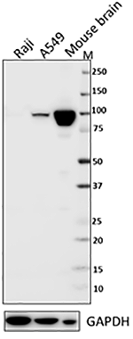
Hela cell nuclear extracts were resolved by electrophoresis, transferred to nitrocellulose and probed with rabbit polyclonal anti-Lamin A/C. Proteins were visualized using a donkey anti-rabbit secondary antibody conjugated to HRP and a chemiluminescence system. -

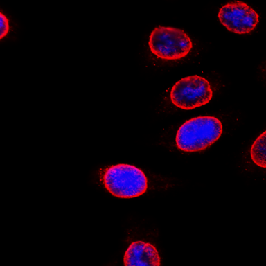
HeLa cells were fixed with 2% PFA for ten minutes, permeabilized with 0.5% Triton X-100 for five minutes, and blocked with 5% FBS for 30 minutes. Then the cells were intracellularly stained with 2 µg/mL anti-Lamin A/C Antibody (clone Poly6135) followed by Alexa Fluor® 594 (red) conjugated goat anti-Rabbit IgG in blocking buffer for two hours at room temperature. Actin filaments were labeled with Alexa Fluor® 488 Phalloidin (green). Nuclei were counterstained with DAPI (blue). The image was captured with a 60X objective.
Lamins A and C are structural components of the lamina, a scaffold of proteins localized to the nuclear inner membrane, and contribute to nuclear membrane dynamics and chromatin structure. Mutations in the LMNA gene have been linked to multiple laminopathy disorders, including Hutchinson-Gilford progeria syndrome (HGPS), an autosomal dominant disorder characterized by the appearance of accelerated and dramatic aging. Patients with HGPS have a mutation in the LMNA gene that results in aberrant splicing and production of the toxic, truncated Lamin A protein progerin that functions in a dominant negative pattern.
Product DetailsProduct Details
- Verified Reactivity
- Human
- Reported Reactivity
- Mouse
- Antibody Type
- Polyclonal
- Host Species
- Rabbit
- Immunogen
- Recombinant (partial), C-terminal
- Formulation
- This antibody is provided in phosphate-buffered solution, pH 7.2, containing 0.09% sodium azide and 50% glycerol.
- Preparation
- The antibody was purified by antigen-affinity chromatography.
- Concentration
- Lot-specific (to obtain lot-specific concentration and expiration, please enter the lot number in our Certificate of Analysis online tool.)
- Storage & Handling
- Upon receipt, store frozen at -20°C.
- Application
-
WB - Quality tested
ICC - Verified - Recommended Usage
-
Each lot of this antibody is quality control tested by Western blotting. Western blotting, suggested working dilution(s): Use 10 µl per 5 ml antibody dilution buffer for each mini-gel. For immunocytochemistry, the suggested usage is 1.0 - 5.0 µg per mL. It is recommended that the reagent be titrated for optimal performance for each application.
- Application Notes
-
This product may contain other non-IgG subtypes
*This antibody weakly cross-reacts with Mouse. -
Application References
(PubMed link indicates BioLegend citation) - Product Citations
-
- RRID
-
AB_315796 (BioLegend Cat. No. 613501)
AB_315797 (BioLegend Cat. No. 613502)
Antigen Details
- Structure
- Intermediate filament family, farnesyl binding domain, dimer. Isoforms A, AD10, C, approximately 70 kD, 66 kD, and 61 kD, respectively
- Distribution
-
Nucleoplasmic side of inner nuclear membrane
- Function
- Fibrous component of nuclear lamina, provides framework for nuclear envelope, may interact with chromatin
- Interaction
- Homodimeric with Lamin C, interacts with LAPs 1A-1C, emerin, Narf, hsMOK2, PKC, SREBP1a, SREBP 1c
- Modification
- Phosphorylation, Methylation, Farnesylation
- Biology Area
- Cell Biology, Cell Motility/Cytoskeleton/Structure, Neuroscience, Neuroscience Cell Markers
- Molecular Family
- Nuclear Markers
- Antigen References
-
1. Machiels B, et al. 1996. J. Biol. Chem. 271:9249.
2. Barton R, et al. 1999. J. Biol. Chem. 274:30008.
3. Gruenbaum Y, et al. 2000. J. Struct. Biol. 129:313.
4. Stierle V, et al. 2003. Biochemistry 42:4819. - Regulation
- Strictly under cell cycle control as seen by disintegration/formation of nuclear envelope in prophase/telophase. Phosphorylation regulates disassembly
- Gene ID
- 4000 View all products for this Gene ID
- UniProt
- View information about Lamin A/C on UniProt.org

Related FAQs
Other Formats
View All Lamin A/C Reagents Request Custom Conjugation| Description | Clone | Applications |
|---|---|---|
| Purified anti-Lamin A/C | Poly6135 | WB,ICC |
Customers Also Purchased
Compare Data Across All Formats
This data display is provided for general comparisons between formats.
Your actual data may vary due to variations in samples, target cells, instruments and their settings, staining conditions, and other factors.
If you need assistance with selecting the best format contact our expert technical support team.


Follow Us