 Login / Register


- Clone
- 259D (See other available formats)
- Regulatory Status
- RUO
- Other Names
- Forkhead box protein P3, Scurfin, JM2, IPEX, Zinc finger protein JM2
- Isotype
- Mouse IgG1, κ
- Ave. Rating
- Submit a Review
- Product Citations
- publications

-

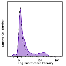
Cell extract from HEK293T cells transfected with human FoxP3 cDNA was resolved by electrophoresis, transferred to nitrocellulose, and probed with monoclonal anti-FoxP3 antibody (clone 259D). Proteins were visualized using a goat anti-mouse secondary conjugated to HRP and a chemiluminescence detection system. -

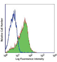
Formalin-fixed, paraffin-embedded Cynomolgus kidney was treated with EDTA pH 8.0 using a high pressure cooker prior to staining. Staining was carried out with monoclonal anti-FoxP3 (clone 259D) at 10 µg/ml followed by biotinylated goat anti-mouse and streptavidin HRP. Staining was visualized with DAB substrate and the tissue counterstained with hematoxylin. -

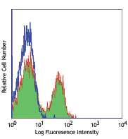
SeqIF™ (sequential immunofluorescence) staining on COMET™ of Purified anti-FOXP3 (clone 259D, yellow) on formalin-fixed paraffin-embedded human head and neck squamous cell carcinoma (HNSCC) tissue at 10 µg/mL. Alexa Fluor™ Plus 647 Goat anti-Mouse IgG antibody (Lunaphore, Cat. No. DR647MS) was used as a secondary antibody. Nuclei were counterstained with DAPI (blue). Tissue underwent an all-in-one dewaxing and antigen retrieval preprocessing.
| Cat # | Size | Price | Quantity Check Availability | Save | ||
|---|---|---|---|---|---|---|
| 320201 | 25 µg | 104€ | ||||
| 320202 | 100 µg | 212€ | ||||
FOXP3 is a 50-55 kD transcription factor, also known as Forkhead box protein P3, Scurfin, JM2, or IPEX. It is proposed to be a master regulatory gene and more specific marker of T regulatory cells than most cell surface markers (such as CD4 and CD25). Transduced expression of FOXP3 in CD4+/CD25- cells has been shown to induce GITR, CD103, and CTLA4 and impart a T regulatory cell phenotype. FOXP3 is mutated in X-linked autoimmunity-allergic dysregulation syndrome (XLAAD or IPEX) in humans and in "scurfy" mice. Overexpression of FOXP3 has been shown to lead to a hypoactive immune state suggesting that this transcriptional factor is a central regulator of T cell activity. In human, unlike in mouse, two isoforms of FOXP3 have been reported: one (FOXP3) corresponding to the canonical full-length sequence; the other (FOXP3 δ2) lacking exon 2. The 259D antibody recognizes human FOXP3 epitope in the region of amino acids 105-235.
Product DetailsProduct Details
- Verified Reactivity
- Human
- Reported Reactivity
- Cynomolgus, Rhesus, Baboon, Chimpanzee
- Antibody Type
- Monoclonal
- Host Species
- Mouse
- Immunogen
- Full-length FOXP3 protein
- Formulation
- This antibody is provided in phosphate-buffered solution, pH 7.2, containing 0.09% sodium azide.
- Preparation
- The antibody was purified by affinity chromatography.
- Concentration
- 0.5 mg/ml
- Storage & Handling
- The antibody solution should be stored undiluted between 2°C and 8°C.
- Application
-
WB - Quality tested
IHC-P - Verified
ICFC - Reported in the literature, not verified in house
SB - Community verified - Recommended Usage
-
Each lot of this antibody is quality control tested by immunofluorescent intracellular staining with flow cytometric analysis. For flow cytometric staining, the suggested use of this reagent is ≤ 0.5 µg per 106 cells in 100 µl volume. For Western blotting, the suggested working dilution(s) is ≤ 5.0 µg/ml in antibody dilution buffer. It is recommended that the reagent be titrated for optimal performance for each application.
- Application Notes
-
Additional reported applications (for the relevant formats) include: Western blotting1, and immunohistochemical staining1 of acetone-fixed frozen sections and formalin-fixed paraffin-embedded sections. The 259D antibody gives strong positivity on paraffin and frozen sections and the antibody stains some epithelial cells. The binding of 206D to FOXP3 can be partially blocked by 259D, but 206D does not show significant blocking effect on 259D binding.
NOTE: For flow cytometric staining with this clone, True-Nuclear™ Transcription Factor Buffer Set (Cat. No. 424401) offers improved staining and is highly recommended. - Additional Product Notes
-
For the use of this antibody in spatial biology applications, we have partnered with Lunaphore Technologies for demonstration of our antibodies on the COMET™. The COMET™ platform is an automated, end-to-end spatial biology solution developed for rapid and flexible multiplex tissue profiling. More information on the COMET™ and a complete list of our antibodies that have been demonstrated on the COMET™ can be found here.
-
Application References
(PubMed link indicates BioLegend citation) -
- Roncador G, et al. 2005 Eur. J. Immunol. 35:1681.
- Yang ZZ, et al. 2006. Blood 107:3639. PubMed
- Gavin MA, et al. 2006. P. Natl. Acad. Sci. USA 103:6659. PubMed
- Groh V, et al. 2006. Nature Immunology 7:755. PubMed
- Tran DQ, et al. 2007. Blood doi:10.1182/blood-2007-06-094656.PubMEd
- Long SA, et al. 2008. J Autoimmun. 30:293. PubMed
- Gong G, et al. 2009. Blood 113:837. PubMed
- Long SA, et al. 2009. Eur J. Immunol. 39:612. PubMed
- Long SA, et al. 2010. Diabetes. 59:407. PubMed
- Ferraro A, et al. 2014. PNAS. 111:1111. PubMed
- Vudattu NK, et al. 2014. J Immunol. 193:587. PubMed
- Dupont G, et al. 2014. Cytokine. 69:146. PubMed
- Product Citations
-
- RRID
-
AB_430884 (BioLegend Cat. No. 320201)
AB_430885 (BioLegend Cat. No. 320202)
Antigen Details
- Structure
- Forkhead/winged-helix transcription factor family, approximately 50 kD, contains zinc finger and forkhead domains
- Distribution
-
Nuclear; expressed in T regulatory cells
- Function
- Transcription factor proposed to be a master regulatory gene in T regulatory cell development and a critical factor for immune homeostasis
- Interaction
- Interacts with DNA
- Cell Type
- Tregs
- Biology Area
- Cell Biology, Immunology, Transcription Factors
- Molecular Family
- Nuclear Markers
- Antigen References
-
1. Hori S, et al. 2003. Science 299:1057.
- Regulation
- FOXP3 is present at high levels in T regulatory cell can also be induced by T cell activation
- Gene ID
- 50943 View all products for this Gene ID
- UniProt
- View information about FOXP3 on UniProt.org

Related FAQs
- Can I stain whole blood with anti-FOXP3 using your Foxp3 staining kit?
-
It is not recommended. It is best to use PBMCs for this testing.
- Can FOXP3 be costained with cytokines?
-
The larger holes created by the nuclear permeabilization required for FOXP3 may allow cytokines to leak out of the cell, making it harder to detect lowly-expressed cytokines. You may have to use a control where the cells are only permeabilized through the cell membrane.
- If an antibody clone has been previously successfully used in IBEX in one fluorescent format, will other antibody formats work as well?
-
It’s likely that other fluorophore conjugates to the same antibody clone will also be compatible with IBEX using the same sample fixation procedure. Ultimately a directly conjugated antibody’s utility in fluorescent imaging and IBEX may be specific to the sample and microscope being used in the experiment. Some antibody clone conjugates may perform better than others due to performance differences in non-specific binding, fluorophore brightness, and other biochemical properties unique to that conjugate.
- Will antibodies my lab is already using for fluorescent or chromogenic IHC work in IBEX?
-
Fundamentally, IBEX as a technique that works much in the same way as single antibody panels or single marker IF/IHC. If you’re already successfully using an antibody clone on a sample of interest, it is likely that clone will have utility in IBEX. It is expected some optimization and testing of different antibody fluorophore conjugates will be required to find a suitable format; however, legacy microscopy techniques like chromogenic IHC on fixed or frozen tissue is an excellent place to start looking for useful antibodies.
- Are other fluorophores compatible with IBEX?
-
Over 18 fluorescent formats have been screened for use in IBEX, however, it is likely that other fluorophores are able to be rapidly bleached in IBEX. If a fluorophore format is already suitable for your imaging platform it can be tested for compatibility in IBEX.
- The same antibody works in one tissue type but not another. What is happening?
-
Differences in tissue properties may impact both the ability of an antibody to bind its target specifically and impact the ability of a specific fluorophore conjugate to overcome the background fluorescent signal in a given tissue. Secondary stains, as well as testing multiple fluorescent conjugates of the same clone, may help to troubleshoot challenging targets or tissues. Using a reference control tissue may also give confidence in the specificity of your staining.
- How can I be sure the staining I’m seeing in my tissue is real?
-
In general, best practices for validating an antibody in traditional chromogenic or fluorescent IHC are applicable to IBEX. Please reference the Nature Methods review on antibody based multiplexed imaging for resources on validating antibodies for IBEX.
Other Formats
View All FOXP3 Reagents Request Custom Conjugation| Description | Clone | Applications |
|---|---|---|
| PE anti-human FOXP3 | 259D | ICFC |
| Purified anti-human FOXP3 | 259D | WB,IHC-P,ICFC,SB |
| Alexa Fluor® 488 anti-human FOXP3 | 259D | ICFC |
| Alexa Fluor® 647 anti-human FOXP3 | 259D | ICFC |
| Pacific Blue™ anti-human FOXP3 | 259D | ICFC |
| True-Nuclear™ Human Treg Flow™ Kit (FOXP3 Alexa Fluor® 488/CD4 PE-Cy5/CD25 PE) | 259D | ICFC |
Customers Also Purchased
Compare Data Across All Formats
This data display is provided for general comparisons between formats.
Your actual data may vary due to variations in samples, target cells, instruments and their settings, staining conditions, and other factors.
If you need assistance with selecting the best format contact our expert technical support team.
-
PE anti-human FOXP3

Human peripheral blood lymphocytes were surface stained with... 
-
Purified anti-human FOXP3

Cell extract from HEK293T cells transfected with human FoxP3... 
Formalin-fixed, paraffin-embedded Cynomolgus kidney was trea... 
SeqIF™ (sequential immunofluorescence) staining on COMET™ of... -
Alexa Fluor® 488 anti-human FOXP3

Human peripheral blood lymphocytes were surface stained with... 
-
Alexa Fluor® 647 anti-human FOXP3

Human peripheral blood lymphocytes were surface stained with... 
-
Pacific Blue™ anti-human FOXP3

Human peripheral blood lymphocytes were surface stained with... 
-
True-Nuclear™ Human Treg Flow™ Kit (FOXP3 Alexa Fluor® 488/CD4 PE-Cy5/CD25 PE)

Human peripheral blood lymphocytes were stained with True-Nu... 


Follow Us