 Login / Register


- Clone
- 3E7/C3b (See other available formats)
- Regulatory Status
- RUO
- Other Names
- Complement Component 3, C3b, iC3b, C3bi
- Isotype
- Mouse IgG1, κ
- Ave. Rating
- Submit a Review
- Product Citations
- publications

-

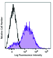
Human peripheral blood cells were opsonized with complement by incubation with purified anti-CD3 (top) or purified isotype control (bottom) and then incubated with fresh human serum (as a source of complement), followed by staining with C3b/iC3b (clone 3E7/C3b) FITC (filled histogram) or mouse IgG1, κ FITC isotype control (open histogram). Data shown was gated in the lymphocyte population. -

| Cat # | Size | Price | Quantity Check Availability | Save | ||
|---|---|---|---|---|---|---|
| 846107 | 25 tests | 81€ | ||||
| 846108 | 100 tests | 203€ | ||||
Complement C3 is a 185 kD glycoprotein composed of two chains linked by a disulfide bond. It is the fourth complement component to react in the classical pathway. It is also a key protein in both the alternative and the lectin complement activation pathways. C3b is proteolytically generated from C3 by cleavage of the C3a-C3b peptide bond in the protein by C3 convertase. C3b can bind covalently, via its reactive thioester, to cell surface carbohydrates or immune aggregates. Macrophages and neutrophils recognize C3b by the complement receptor 1 (CR1, CD35). Opsonization of target surfaces with C3b is central to all three pathways of complement activation. The proteolytically inactive forms of C3b, iC3b, are generated by cleavage of the alpha chain at one or two positions by factor I in the presence of co-factors, such as factor H. A fragment of C3b, C3f, with a molecular weight of approximately 2 kD is generated when C3b is cleaved at two positions by factor I. Although iC3b is less active than C3b, iC3b’s interactions with lymphoid and phagocytic cells via CR2 (CD21) and CR3 (CD11b/CD18) can expand target-specific B and T cells. iC3b can be cleaved to form C3c and C3dg.
Product DetailsProduct Details
- Verified Reactivity
- Human
- Reported Reactivity
- Non-Human Primate
- Antibody Type
- Monoclonal
- Host Species
- Mouse
- Immunogen
- Purified native human C3b protein
- Formulation
- Phosphate-buffered solution, pH 7.2, containing 0.09% sodium azide and BSA (origin USA)
- Preparation
- The antibody was purified by affinity chromatography and conjugated with FITC under optimal conditions.
- Concentration
- Lot-specific (to obtain lot-specific concentration and expiration, please enter the lot number in our Certificate of Analysis online tool.)
- Storage & Handling
- The antibody solution should be stored undiluted between 2°C and 8°C, and protected from prolonged exposure to light. Do not freeze.
- Application
-
FC - Quality tested
- Recommended Usage
-
Each lot of this antibody is quality control tested by immunofluorescent staining with flow cytometric analysis. For flow cytometric staining, the suggested use of this reagent is 5 µl per million cells in 100 µl staining volume or 5 µl per 100 µl of whole blood.
- Excitation Laser
-
Blue Laser (488 nm)
- Application Notes
-
Additional reported applications (for the relevant formats) include: Blocking3, ELISA Detection1, immunofluorescent staining1,2, and RIA2,5.
This antibody recognizes C3, C3b and iC3b, and does not cross-react with C3d.
-
Application References
(PubMed link indicates BioLegend citation) -
- Pokrass M, et al. 2013. Mol. Immunol. 56:549. (ELISA, IF)
- Kennedy AD, et al. 2003. Blood. 101:1071. (FC, IF, RIA)
- DiLillo DJ, et al. 2006. Mol. Immunol. 43:1010-9.
- Lindorfer MA, et al. 2010 Blood 115:2283. (Block)
- Sokoloff MH, et al. 2000. Cancer Immunol. Immunother. 49:551. (RIA, FC)
- Product Citations
-
- RRID
-
AB_2632796 (BioLegend Cat. No. 846107)
AB_2632797 (BioLegend Cat. No. 846108)
Antigen Details
- Structure
- C3b is a glycosylated, 176 kD protein that is formed by the cleavage of C3. iC3b is proteolytically derived from C3b and exists as a mixture of 176 and 174 kD proteins.
- Distribution
-
Extracellular fluid, plasma membrane, and serum.
- Function
- C3b is involved in all three complement pathways and is essential for effective complement activation and presentation of antigens to cells of the adaptive immune system. iC3b is less active than C3b, but the target-bound protein can expand target-specific B-cell and T-cell populations by interacting with lymphoid and phagocytic cells.
- Interaction
- C3b can bind covalently, via its reactive thioester, to cell surface carbohydrates or immune aggregates. Macrophages and neutrophils have receptors, CR1 (CD35), for C3b. iC3b can also function as opsonin, but it binds through CR2 and CR3.
- Ligand/Receptor
- C3b can bind to Factor B, Factor P, Factor H, Factor I, C5, DAF (CD55), MCP (CD46), and CR1 (CD35). iC3b can interact with CR2 (CD21), and CR3 (CD11b/CD18).
- Biology Area
- Cell Biology, Complement, Immunology, Innate Immunity, Neuroinflammation, Neuroscience
- Antigen References
-
1. Lambris JD. 1988. Immunol. Today 9:387-93.
- Gene ID
- 718 View all products for this Gene ID
- UniProt
- View information about C3b/iC3b on UniProt.org


Related FAQs
Other Formats
View All C3b/iC3b Reagents Request Custom Conjugation| Description | Clone | Applications |
|---|---|---|
| Purified anti-complement C3b/iC3b | 3E7/C3b | FC,Direct ELISA,Block,ELISA Detection,ICC,RIA |
| FITC anti-complement C3b/iC3b | 3E7/C3b | FC |
| APC anti-complement C3b/iC3b | 3E7/C3b | FC |
| PE anti-complement C3b/iC3b | 3E7/C3b | FC |
Customers Also Purchased

Compare Data Across All Formats
This data display is provided for general comparisons between formats.
Your actual data may vary due to variations in samples, target cells, instruments and their settings, staining conditions, and other factors.
If you need assistance with selecting the best format contact our expert technical support team.
-
Purified anti-complement C3b/iC3b

Human peripheral mononucleated cells were incubated with pur... -
FITC anti-complement C3b/iC3b

Human peripheral blood cells were opsonized with complement ... 
-
APC anti-complement C3b/iC3b

Human peripheral blood cells were opsonized with complement ... 
-
PE anti-complement C3b/iC3b

Human peripheral blood cells were opsonized with complement ... 


Follow Us