 Login / Register


- Clone
- RM4-4 (See other available formats)
- Regulatory Status
- RUO
- Other Names
- L3T4, T4
- Isotype
- Rat IgG2b, κ
- Ave. Rating
- Submit a Review
- Product Citations
- publications

-

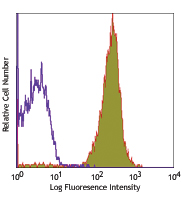
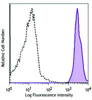
C57BL/6 mouse splenocytes were stained with CD3 APC and anti-mouse CD4 (clone RM4-4) Brilliant Violet 605™ (left) or Rat IgG2b, κ Isotype Control (right).
| Cat # | Size | Price | Quantity Check Availability | Save | ||
|---|---|---|---|---|---|---|
| 116027 | 50 µg | 248€ | ||||
CD4 is a 55 kD protein, also known as L3T4 or T4. It is a member of the Ig superfamily, primarily expressed on most thymocytes and a subset of T cells, and weakly on macrophages and dendritic cells. It acts as a coreceptor with the TCR during T cell activation and thymic differentiation by binding MHC class II and associating with the protein tyrosin kinase, lck.
Product DetailsProduct Details
- Verified Reactivity
- Mouse
- Antibody Type
- Monoclonal
- Host Species
- Rat
- Immunogen
- BALB/c mouse thymocytes
- Formulation
- Phosphate-buffered solution, pH 7.2, containing 0.09% sodium azide and BSA (origin USA).
- Preparation
- The antibody was purified by affinity chromatography and conjugated with Brilliant Violet 605™ under optimal conditions.
- Concentration
- 0.2 mg/ml
- Storage & Handling
- The antibody solution should be stored undiluted between 2°C and 8°C, and protected from prolonged exposure to light. Do not freeze.
- Application
-
FC - Quality tested
- Recommended Usage
-
Each lot of this antibody is quality control tested by immunofluorescent staining with flow cytometric analysis. For flow cytometric staining, the suggested use of this reagent is ≤ 0.25 µg per million cells in 100 µl volume. It is recommended that the reagent be titrated for optimal performance for each application.
Brilliant Violet 605™ excites at 405 nm and emits at 603 nm. The bandpass filter 610/20 nm is recommended for detection, although filter optimization may be required depending on other fluorophores used. Be sure to verify that your cytometer configuration and software setup are appropriate for detecting this channel. Refer to your instrument manual or manufacturer for support. Brilliant Violet 605™ is a trademark of Sirigen Group Ltd.
Learn more about Brilliant Violet™.
This product is subject to proprietary rights of Sirigen Inc. and is made and sold under license from Sirigen Inc. The purchase of this product conveys to the buyer a non-transferable right to use the purchased product for research purposes only. This product may not be resold or incorporated in any manner into another product for resale. Any use for therapeutics or diagnostics is strictly prohibited. This product is covered by U.S. Patent(s), pending patent applications and foreign equivalents. - Excitation Laser
-
Violet Laser (405 nm)
- Application Notes
-
RM4-4 antibody does not block the binding of GK1.5 and RM4-5 antibodies to CD4 T cells. For immunohistochemistry applications, the RM4-5 (Cat. No. 100506) and GK1.5 (Cat. No. 100402) antibodies are recommended.
-
Application References
(PubMed link indicates BioLegend citation) -
- Bendelac A. 1995. Curr. Opin. Immunol. 7:367.
- Norian LA and Allen PM. 2004. J. Immunol. 173:835.
- Product Citations
-
- RRID
-
AB_2800581 (BioLegend Cat. No. 116027)
Antigen Details
- Structure
- Ig superfamily, 55 kD
- Distribution
-
Majority of thymocytes, T cell subset
- Function
- TCR co-receptor, T cell activation
- Ligand/Receptor
- MHC class II molecule
- Cell Type
- T cells, Thymocytes, Tregs
- Biology Area
- Immunology
- Molecular Family
- CD Molecules
- Antigen References
-
1. Barclay A, et al. 1997. The Leukocyte Antigen FactsBook Academic Press.
2. Bierer BE, et al. 1989. Annu. Rev. Immunol. 7:579.
3. Janeway CA. 1992. Annu. Rev. Immunol. 10:645. - Gene ID
- 12504 View all products for this Gene ID
- UniProt
- View information about CD4 on UniProt.org
Related Pages & Pathways
Pathways


Related FAQs
- I am unable to see expression of T cell markers such as CD3 and CD4 post activation.
- TCR-CD3 complexes on the T-lymphocyte surface are rapidly downregulated upon activation with peptide-MHC complex, superantigen or cross-linking with anti-TCR or anti-CD3 antibodies. PMA/Ionomycin treatment has been shown to downregulate surface CD4 expression. Receptor downregulation is a common biological phenomenon and so make sure that your stimulation treatment is not causing it in your sample type.
Other Formats
View All CD4 Reagents Request Custom Conjugation| Description | Clone | Applications |
|---|---|---|
| FITC anti-mouse CD4 | RM4-4 | FC |
| PE anti-mouse CD4 | RM4-4 | FC |
| Pacific Blue™ anti-mouse CD4 | RM4-4 | FC |
| Biotin anti-mouse CD4 | RM4-4 | FC |
| PerCP/Cyanine5.5 anti-mouse CD4 | RM4-4 | FC |
| APC anti-mouse CD4 | RM4-4 | FC |
| PE/Cyanine7 anti-mouse CD4 | RM4-4 | FC |
| Purified anti-mouse CD4 | RM4-4 | FC |
| APC/Fire™ 750 anti-mouse CD4 | RM4-4 | FC |
| Alexa Fluor® 700 anti-mouse CD4 | RM4-4 | FC |
| Brilliant Violet 510™ anti-mouse CD4 | RM4-4 | FC |
| Brilliant Violet 421™ anti-mouse CD4 | RM4-4 | FC |
| Brilliant Violet 605™ anti-mouse CD4 | RM4-4 | FC |
Customers Also Purchased
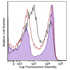
Compare Data Across All Formats
This data display is provided for general comparisons between formats.
Your actual data may vary due to variations in samples, target cells, instruments and their settings, staining conditions, and other factors.
If you need assistance with selecting the best format contact our expert technical support team.
-
FITC anti-mouse CD4

C57BL/6 mouse splenocytes stained with RM4-4 FITC -
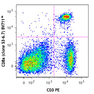
PE anti-mouse CD4

C57BL/6 mouse splenocytes stained with CD3 (145-2C11) FITC a... -
Pacific Blue™ anti-mouse CD4

C57BL/6 mouse splenocytes were stained with CD4 (clone RM4-4... -
Biotin anti-mouse CD4

C57BL/6 mouse splenocytes were stained with biotinylated CD4... -
PerCP/Cyanine5.5 anti-mouse CD4

C57BL/6 mouse splenocytes were stained with CD3e FITC and CD... -
APC anti-mouse CD4

C57BL/6 mouse splenocytes were stained with CD4 (clone RM4-4... -
PE/Cyanine7 anti-mouse CD4

C57BL/6 mouse splenocytes were stained with CD4 (clone RM4-4... -
Purified anti-mouse CD4

C57BL/6 mouse splenocytes stained with purified CD4 (clone R... -
APC/Fire™ 750 anti-mouse CD4

C57BL/6 mouse splenocytes stained with CD3 (145-2C11) PE and... 
-
Alexa Fluor® 700 anti-mouse CD4

C57BL/6 mouse splenocytes stained with CD3 (145-2C11) PE and... 
-
Brilliant Violet 510™ anti-mouse CD4

C57BL/6 mouse splenocytes were stained with CD3 FITC and CD4... -
Brilliant Violet 421™ anti-mouse CD4

C57BL/6 mouse splenocytes were stained with CD3 FITC and CD4... -
Brilliant Violet 605™ anti-mouse CD4

C57BL/6 mouse splenocytes were stained with CD3 APC and anti...


Follow Us