 Login / Register


- Clone
- 3.9 (See other available formats)
- Regulatory Status
- RUO
- Workshop
- III NL707
- Other Names
- Integrin αX subunit, CR4, p150, ITGAX
- Isotype
- Mouse IgG1, κ
- Ave. Rating
- Submit a Review
- Product Citations
- publications

-

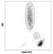
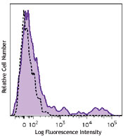
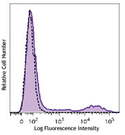
Human peripheral blood granulocytes were stained with CD11c (clone 3.9) Brilliant Violet 510™ (filled histogram) or mouse IgG1, κ Brilliant Violet 510™ isotype control (open histogram). -

Human peripheral blood mononuclear cell (PBMC) derived dendritic cells were fixed with 1% paraformaldehyde (PFA), and then stained with 10 µg/ml of CD11c (clone 3.9) Brilliant Violet 510™ (yellow) for 30 minutes at room temperature. Nuclei were counterstained with DAPI (blue). The image was captured by 40X objective.
| Cat # | Size | Price | Quantity Check Availability | Save | ||
|---|---|---|---|---|---|---|
| 301633 | 25 tests | 146€ | ||||
| 301634 | 100 tests | 340€ | ||||
CD11c is a 145-150 kD type I transmembrane glycoprotein also known as integrin αX and CR4. CD11c non-covalently associates with integrin β2 (CD18) and is expressed on monocytes/macrophages, dendritic cells, granulocytes, NK cells, and subsets of T and B cells. CD11c has been reported to play a role in adhesion and CTL killing through its interactions with fibrinogen, CD54, and iC3b.
Product DetailsProduct Details
- Verified Reactivity
- Human, Cynomolgus, Rhesus
- Reported Reactivity
- African Green, Baboon, Chimpanzee, Squirrel Monkey
- Antibody Type
- Monoclonal
- Host Species
- Mouse
- Formulation
- Phosphate-buffered solution, pH 7.2, containing 0.09% sodium azide and BSA (origin USA).
- Preparation
- The antibody was purified by affinity chromatography and conjugated with Brilliant Violet 510™ under optimal conditions.
- Concentration
- Lot-specific (to obtain lot-specific concentration and expiration, please enter the lot number in our Certificate of Analysis online tool.)
- Storage & Handling
- The antibody solution should be stored undiluted between 2°C and 8°C, and protected from prolonged exposure to light. Do not freeze.
- Application
-
FC - Quality tested
ICC - Verified - Recommended Usage
-
Each lot of this antibody is quality control tested by immunofluorescent staining with flow cytometric analysis. For flow cytometric staining, the suggested use of this reagent is 5 µL per million cells in 100 µL staining volume or 5 µL per 100 µL of whole blood. It is recommended that the reagent be titrated for optimal performance for each application.
Brilliant Violet 510™ excites at 405 nm and emits at 510 nm. The bandpass filter 510/50 nm is recommended for detection, although filter optimization may be required depending on other fluorophores used. Be sure to verify that your cytometer configuration and software setup are appropriate for detecting this channel. Refer to your instrument manual or manufacturer for support. Brilliant Violet 510™ is a trademark of Sirigen Group Ltd.
Learn more about Brilliant Violet™.
This product is subject to proprietary rights of Sirigen Inc. and is made and sold under license from Sirigen Inc. The purchase of this product conveys to the buyer a non-transferable right to use the purchased product for research purposes only. This product may not be resold or incorporated in any manner into another product for resale. Any use for therapeutics or diagnostics is strictly prohibited. This product is covered by U.S. Patent(s), pending patent applications and foreign equivalents. - Excitation Laser
-
Violet Laser (405 nm)
- Application Notes
-
Clone 3.9 preferentially binds the activated form of CD11c, is specific for the I domain of CD11c, and is able to partially block the binding of CD11c and ICAM-4. 3.9 binding is divalent cation dependent12. While analyzing blood, it is best to use heparin as the anti-coagulant and not EDTA. Since the ability of clone 3.9 to bind to its target is divalent cation dependent, the usage of EDTA as an anti-coagulant may be detrimental to staining due to its chelating properties.
Additional reported applications (for the relevant formats) include: immunohistochemical staining of acetone-fixed frozen tissue sections4, and functional assays5,6. The LEAF™ purified antibody (Endotoxin <0.1 EU/μg, Azide-Free, 0.2 μm filtered) is recommended for functional assays (Cat. No. 301616). For highly sensitive assays, we recommend Ultra-LEAF™ purified antibody (Cat. No. 301632) with a lower endotoxin limit than standard LEAF™ purified antibodies (Endotoxin <0.01 EU/µg). -
Application References
(PubMed link indicates BioLegend citation) -
- Schlossman S, et al. Eds. 1995. Leucocyte Typing V. Oxford University Press. New York.
- Knapp W, et al. 1989. Leucocyte Typing IV Oxford University Press. New York.
- McMichael A, et al. Eds. 1987. Leucocyte Typing III Oxford University Press. New York.
- Vainer B, et al. 2000. Am. J. Surg. Pathol. 24:1115. (IHC)
- Ottonello L, et al. 1999. Blood 93:3505.
- Metelitsa LS, et al. 2002. Blood 99:4166.
- Sadhu C, et al. 2007. J. Leukoc. Biol. doi:10.1189/jlb.1106680. PubMed
- Ihanus E, et al. 2007. Blood 109:802-810.
- Gurer C, et al. 2008. Blood 112:1231. PubMed
- Asai A, et al. 2009. J. Lipid Res. 50:95. PubMed
- Yoshino N, et al. 2000. Exp. Anim. (Tokyo) 49:97. (FC)
- Sadhu C, et al. 2008. J. Immunoass. Immunoch. 29:42. (FC)
- Product Citations
-
- RRID
-
AB_2561990 (BioLegend Cat. No. 301633)
AB_2563795 (BioLegend Cat. No. 301634)
Antigen Details
- Structure
- Integrin, type I transmembrane glycoprotein, associates with integrin β2 (CD18), 145-150 kD
- Distribution
-
Myeloid, dendritic cells, NK cells, B cells and T cell subsets
- Function
- Adhesion, CTL killing
- Ligand/Receptor
- CD54, fibrinogen, iC3b, ICAM-1, ICAM-4
- Cell Type
- B cells, Dendritic cells, Neutrophils, NK cells, T cells, Tregs
- Biology Area
- Cell Adhesion, Cell Biology, Costimulatory Molecules, Immunology, Innate Immunity, Neuroscience, Neuroscience Cell Markers
- Molecular Family
- Adhesion Molecules, CD Molecules
- Antigen References
-
1. Petty H. 1996. Immunol. Today 17:209.
2. Springer T. 1994. Cell 76:301.
3. Ihanus E, et al. 2007. Blood 109:802-810. - Gene ID
- 3687 View all products for this Gene ID
- UniProt
- View information about CD11c on UniProt.org
Related Pages & Pathways
Pathways


Related FAQs
Other Formats
View All CD11c Reagents Request Custom Conjugation| Description | Clone | Applications |
|---|---|---|
| FITC anti-human CD11c | 3.9 | FC |
| PE anti-human CD11c | 3.9 | FC |
| Purified anti-human CD11c | 3.9 | FC,CyTOF®,IHC |
| PE/Cyanine7 anti-human CD11c | 3.9 | FC |
| PE/Cyanine5 anti-human CD11c | 3.9 | FC |
| Biotin anti-human CD11c | 3.9 | FC |
| APC anti-human CD11c | 3.9 | FC |
| Alexa Fluor® 488 anti-human CD11c | 3.9 | FC |
| Alexa Fluor® 647 anti-human CD11c | 3.9 | FC |
| Pacific Blue™ anti-human CD11c | 3.9 | FC |
| PerCP/Cyanine5.5 anti-human CD11c | 3.9 | FC |
| Brilliant Violet 421™ anti-human CD11c | 3.9 | FC |
| Brilliant Violet 711™ anti-human CD11c | 3.9 | FC |
| Ultra-LEAF™ Purified anti-human CD11c | 3.9 | FC,CyTOF®,Block,IHC |
| Brilliant Violet 510™ anti-human CD11c | 3.9 | FC,ICC |
| Brilliant Violet 605™ anti-human CD11c | 3.9 | FC |
| Brilliant Violet 650™ anti-human CD11c | 3.9 | FC |
| Purified anti-human CD11c (Maxpar® Ready) | 3.9 | FC,CyTOF® |
| PE/Dazzle™ 594 anti-human CD11c | 3.9 | FC |
| Brilliant Violet 785™ anti-human CD11c | 3.9 | FC |
| Alexa Fluor® 700 anti-human CD11c | 3.9 | FC |
| APC/Fire™ 750 anti-human CD11c | 3.9 | FC |
| Spark Red™ 718 anti-human CD11c | 3.9 | FC |
Customers Also Purchased

Compare Data Across All Formats
This data display is provided for general comparisons between formats.
Your actual data may vary due to variations in samples, target cells, instruments and their settings, staining conditions, and other factors.
If you need assistance with selecting the best format contact our expert technical support team.
-
FITC anti-human CD11c

Human peripheral blood granulocytes were stained with CD11c ... -
PE anti-human CD11c

Human peripheral blood granulocytes stained with 3.9 PE -
Purified anti-human CD11c

Human peripheral blood granulocytes were stained with purifi... -
PE/Cyanine7 anti-human CD11c

Human peripheral blood granulocytes stained with 3.9 PE/Cyan... -
PE/Cyanine5 anti-human CD11c

Human peripheral blood granulocytes stained with 3.9 PE/Cyan... -
Biotin anti-human CD11c

Human peripheral blood granulocytes stained with biotinylate... -
APC anti-human CD11c

Human peripheral blood monocytes stained with 3.9 APC -
Alexa Fluor® 488 anti-human CD11c

Human peripheral blood monocytes stained with 3.9 Alexa Fluo... -
Alexa Fluor® 647 anti-human CD11c

Human peripheral blood monocytes stained with 3.9 Alexa Fluo... -
Pacific Blue™ anti-human CD11c

Human peripheral blood monocytes stained with anti-human CD1... -
PerCP/Cyanine5.5 anti-human CD11c

Human peripheral blood granulocytes were stained with CD11c ... 
Human peripheral blood monocytes were stained with CD11c (cl... -
Brilliant Violet 421™ anti-human CD11c

Human peripheral blood granulocytes were stained with CD11c ... -
Brilliant Violet 711™ anti-human CD11c

Human peripheral blood granulocytes were stained with CD11c ... -
Ultra-LEAF™ Purified anti-human CD11c

Human peripheral blood granulocytes were stained with Ultra-... -
Brilliant Violet 510™ anti-human CD11c

Human peripheral blood granulocytes were stained with CD11c ... 
Human peripheral blood mononuclear cell (PBMC) derived dendr... -
Brilliant Violet 605™ anti-human CD11c

Human peripheral blood granulocytes were stained with CD11c ... -
Brilliant Violet 650™ anti-human CD11c

Human peripheral blood granulocytes were stained with CD11c ... -
Purified anti-human CD11c (Maxpar® Ready)
-
PE/Dazzle™ 594 anti-human CD11c

Human peripheral blood granulocytes were stained with CD11c ... -
Brilliant Violet 785™ anti-human CD11c

Human peripheral blood granulocytes were stained with CD11c ... -
Alexa Fluor® 700 anti-human CD11c

Human peripheral blood monocytes were stained with Alexa Flo... 
Human peripheral blood lymphocytes were stained with PE anti... -
APC/Fire™ 750 anti-human CD11c

Human peripheral blood monocytes were stained with APC/Fire™... 
Human peripheral blood lymphocytes were stained with PE anti... -
Spark Red™ 718 anti-human CD11c

Human peripheral blood monocytes were stained with anti-huma... 
Human peripheral blood lymphocytes were stained with anti-hu...


Follow Us