 Login / Register


- Clone
- 53-7.3 (See other available formats)
- Regulatory Status
- RUO
- Other Names
- Lyt-1, Ly-1, T1, Tp67, Ly-12
- Isotype
- Rat IgG2a, κ
- Ave. Rating
- Submit a Review
- Product Citations
- publications

-

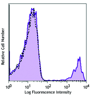
C57BL/6 mouse splenocytes stained with biotinylated 53-7.3, followed by Sav-PE
| Cat # | Size | Price | Quantity Check Availability | Save | ||
|---|---|---|---|---|---|---|
| 100603 | 50 µg | 44€ | ||||
| 100604 | 500 µg | 210€ | ||||
CD5 is a 67 kD protein, also known as Lyt-1, Ly-1, T1, Tp67, or Ly-12. It is a member of the scavenger receptor cysteine-rich protein superfamily (SRCR) and primarily expressed on thymocytes, T cells, and B-1 cells. Although mature α/β T cells express high levels of CD5, very few γ/δ T cells express this antigen. The interaction of CD5 with CD72, gp35-37, TCR, or BCR is involved in T and B cell activation.
Product DetailsProduct Details
- Verified Reactivity
- Mouse
- Antibody Type
- Monoclonal
- Host Species
- Rat
- Immunogen
- Mouse thymus or spleen
- Formulation
- Phosphate-buffered solution, pH 7.2, containing 0.09% sodium azide.
- Preparation
- The antibody was purified by affinity chromatography, and conjugated with biotin under optimal conditions.
- Concentration
- 0.5 mg/ml
- Storage & Handling
- The antibody solution should be stored undiluted between 2°C and 8°C. Do not freeze.
- Application
-
FC - Quality tested
- Recommended Usage
-
Each lot of this antibody is quality control tested by immunofluorescent staining with flow cytometric analysis. For flow cytometric staining, the suggested use of this reagent is ≤0.06 µg per million cells in 100 µl volume. It is recommended that the reagent be titrated for optimal performance for each application.
- Application Notes
-
Additional reported applications (for the relevant formats) include: immunoprecipitation1, and immunohistochemistry2 of acetone-fixed frozen tissue sections, zinc-fixed paraffin-embedded sections and formalin-fixed paraffin-embedded sections.
-
Application References
(PubMed link indicates BioLegend citation) -
- Ledbetter JA, et al. 1979. Immunol. Rev. 47:63. (IP)
- Ledbetter JA, et al. 1980. J. Exp. Med. 152:280. (FC, IHC)
- Bourdeau A, et al. 2007. Blood doi:10.1182/blood-2006-08-044370.
- Product Citations
-
- RRID
-
AB_312732 (BioLegend Cat. No. 100603)
AB_312733 (BioLegend Cat. No. 100604)
Antigen Details
- Structure
- Member of the scavenger receptor cysteine-rich protein superfamily (SRCR), 67 kD
- Distribution
-
Thymocytes, T cells, B-1 cells
- Function
- Negative regulator of T-B cell interaction
- Ligand/Receptor
- CD72, gp35-37
- Cell Type
- B cells, T cells, Thymocytes
- Biology Area
- Costimulatory Molecules, Immunology
- Molecular Family
- CD Molecules
- Antigen References
-
1. Barclay A, et al. 1997. The Leukocyte Antigen FactsBook Academic Press.
2. Kipps TJ. 1988. Adv. Immunol. 47:117.
3. Antin JH, et al. 1985. J. Immunol. 136:505.
4. Tarakhovsky A, et al. 1995. Science 269:535. - Gene ID
- 12507 View all products for this Gene ID
- UniProt
- View information about CD5 on UniProt.org
Related Pages & Pathways
Pathways


Related FAQs
- How many biotin molecules are per antibody structure?
- We don't routinely measure the number of biotins with our antibody products but the number of biotin molecules range from 3-6 molecules per antibody.
Other Formats
View All CD5 Reagents Request Custom ConjugationCustomers Also Purchased
Compare Data Across All Formats
This data display is provided for general comparisons between formats.
Your actual data may vary due to variations in samples, target cells, instruments and their settings, staining conditions, and other factors.
If you need assistance with selecting the best format contact our expert technical support team.
-
Biotin anti-mouse CD5

C57BL/6 mouse splenocytes stained with biotinylated 53-7.3, ... -
FITC anti-mouse CD5

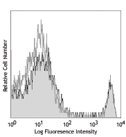
C57BL/6 mouse splenocytes stained with 53-7.3 FITC -
PE anti-mouse CD5

C57BL/6 mouse splenocytes stained with 53-7.3 PE -
PE/Cyanine5 anti-mouse CD5

C57BL/6 mouse splenocytes stained with 53-7.3 PE/Cyanine5 -
Purified anti-mouse CD5

C57BL/6 mouse splenocytes stained with purified 53-7.3, foll... 
Fresh, frozen mouse spleen was stained with purified CD5 clo... -
Brilliant Violet 510™ anti-mouse CD5

C57BL/6 mouse splenocytes were stained with CD5 (clone 53-7.... -
Alexa Fluor® 488 anti-mouse CD5

C57BL/6 mouse splenocytes stained with 53-7.3 Alexa Fluor® 4... -
Alexa Fluor® 647 anti-mouse CD5

C57BL/6 mouse splenocytes stained with 53-7.3 Alexa Fluor® 6... -
PerCP anti-mouse CD5

C57BL/6 mouse splenocytes stained with 53-7.3 PerCP -
Brilliant Violet 421™ anti-mouse CD5

C57BL/6 mouse splenocytes were stained with CD5 (clone 53-7.... -
PE/Cyanine7 anti-mouse CD5

C57BL/6 mouse splenocytes were stained with CD5 (clone 53-7.... -
Purified anti-mouse CD5 (Maxpar® Ready)

C57BL/6 mouse splenocytes (blue) and human Jurkat T cells (r... -
PerCP/Cyanine5.5 anti-mouse CD5

C57BL/6 mouse splenocytes were stained with CD5 (clone 53-7.... -
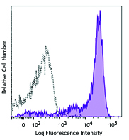
APC anti-mouse CD5

C57BL/6 mouse splenocytes were stained with CD5 (clone 53-7.... -
Alexa Fluor® 594 anti-mouse CD5

C57BL/6 mouse frozen spleen section was fixed with 4% parafo... -
APC/Fire™ 750 anti-mouse CD5

C57BL/6 mouse splenocytes were stained with CD5 (clone 53-7.... -
Alexa Fluor® 700 anti-mouse CD5

C57BL/6 mouse splenocytes were stained with CD5 (clone 53-7.... -
TotalSeq™-A0111 anti-mouse CD5
-
Brilliant Violet 711™ anti-mouse CD5

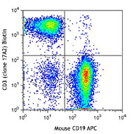
C57BL/6 mouse splenocytes were stained with CD3 FITC and CD5... -
Pacific Blue™ anti-mouse CD5

C57BL/6 mouse splenocytes were stained with CD3 FITC and CD5... -
PE/Dazzle™ 594 anti-mouse CD5

C57BL/6 splenocytes were stained with CD3 APC/Cyanine7 and C... -
TotalSeq™-B0111 anti-mouse CD5
-
TotalSeq™-C0111 anti-mouse CD5
-
APC/Cyanine7 anti-mouse CD5

C57BL/6 mouse splenocytes were stained with CD3 Alexa Fluor®... -
Brilliant Violet 605™ anti-mouse CD5

C57BL/6 mouse splenocytes were stained with anti-mouse CD3 F...


Follow Us