 Login / Register


- Clone
- 4B4-1 (See other available formats)
- Regulatory Status
- RUO
- Workshop
- VI C-7
- Other Names
- 4-1BB, ILA, CD137, TNFRSF9
- Isotype
- Mouse IgG1, κ
- Ave. Rating
- Submit a Review
- Product Citations
- publications

-

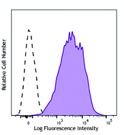
PHA-stimulated (3 days) human peripheral blood mononuclear cells stained with CD137 (clone 4B4-1) APC (filled histogram) or mouse IgG1, κ APC isotype control (open histogram).
| Cat # | Size | Price | Quantity Check Availability | Save | ||
|---|---|---|---|---|---|---|
| 309809 | 25 tests | 116€ | ||||
| 309810 | 100 tests | 266€ | ||||
CD137 is a 39 kD transmembrane protein also known as 4-1BB. It is expressed on activated T cells. CD137 is a type I membrane protein and a member of the tumor necrosis factor receptor superfamily. CD137 appears to be important for T cell proliferation and survival, and induces monocyte activation through its interaction with 4-1BB ligand.
Product DetailsProduct Details
- Verified Reactivity
- Human
- Reported Reactivity
- Chimpanzee, Baboon, Cynomolgus, Rhesus
- Antibody Type
- Monoclonal
- Host Species
- Mouse
- Immunogen
- Ectodomain of recombinant human 4-1BB fusion protein
- Formulation
- Phosphate-buffered solution, pH 7.2, containing 0.09% sodium azide and BSA (origin USA)
- Preparation
- The antibody was purified by affinity chromatography, and conjugated with APC under optimal conditions.
- Concentration
- Lot-specific (to obtain lot-specific concentration and expiration, please enter the lot number in our Certificate of Analysis online tool.)
- Storage & Handling
- The antibody solution should be stored undiluted between 2°C and 8°C, and protected from prolonged exposure to light. Do not freeze.
- Application
-
FC - Quality tested
- Recommended Usage
-
Each lot of this antibody is quality control tested by immunofluorescent staining with flow cytometric analysis. For flow cytometric staining, the suggested use of this reagent is 5 µl per million cells in 100 µl staining volume or 5 µl per 100 µl of whole blood.
- Excitation Laser
-
Red Laser (633 nm)
- Application Notes
-
Additional reported applications (for the relevant formats) include: immunoprecipitation1,4, inhibition of cytokine production2,3, and ELISA. For most successful immunofluorescent staining results, it may be important to maximize signal over background by using a relatively bright fluorochrome-antibody conjugate (Cat. No. 309804) or by using a high sensitivity, three-layer staining technique (e.g., including a biotinylated anti-mouse IgG second step (Cat. No. 405303), followed by Streptavidin-PE (Cat. No. 405204)).
-
Application References
(PubMed link indicates BioLegend citation) -
- Garni-Wagner B, et al. 1996. Cell. Immunol. 169:91. (IP)
- Salih HR, et al. 2000. J. Immunol. 165:2903. (FA)
- Kienzle G, et al. 2000. Int. Immunol. 12:73. (FA)
- Langstein J, et al. 1998. J. Immunol. 160:2488. (IP)
- Product Citations
-
- RRID
-
AB_830671 (BioLegend Cat. No. 309809)
AB_830672 (BioLegend Cat. No. 309810)
Antigen Details
- Structure
- TNFR superfamily, type I transmembrane protein, 30 kD
- Distribution
-
Activated T cells
- Function
- T cell costimulation
- Ligand/Receptor
- 4-1BB ligand
- Cell Type
- T cells
- Biology Area
- Costimulatory Molecules, Immunology
- Molecular Family
- CD Molecules
- Antigen References
-
1. Gruss H, et al. 1995. Blood 85:3378.
2. Sica G, et al. 2000. Adv. Exp. Med. Biol. 465:355.
3. Alderson M, et al. 1994. Eur. J. Immunol. 24:2219.
4. Schwarz H, et al. 1996. Blood 87:2839. - Gene ID
- 3604 View all products for this Gene ID
- UniProt
- View information about CD137 on UniProt.org
Related Pages & Pathways
Pathways


Related FAQs
Other Formats
View All CD137 Reagents Request Custom ConjugationCustomers Also Purchased
Compare Data Across All Formats
This data display is provided for general comparisons between formats.
Your actual data may vary due to variations in samples, target cells, instruments and their settings, staining conditions, and other factors.
If you need assistance with selecting the best format contact our expert technical support team.
-
Purified anti-human CD137 (4-1BB)

PHA-stimulated (3 days) human peripheral blood mononuclear c... -
PE anti-human CD137 (4-1BB)

PHA stimulated (3 days) peripheral blood mononuclear cells s... -
Biotin anti-human CD137 (4-1BB)

PHA-stimulated (4 days) human peripheral blood mononuclear c... -
PE/Cyanine5 anti-human CD137 (4-1BB)

PHA-stimulated (2 days) human peripheral blood mononuclear c... -
APC anti-human CD137 (4-1BB)

PHA-stimulated (3 days) human peripheral blood mononuclear c... -
PerCP/Cyanine5.5 anti-human CD137 (4-1BB)

PHA-stimulated (3 day) human peripheral blood lymphocytes st... -
Alexa Fluor® 700 anti-human CD137 (4-1BB)

PHA-stimulated (3 days) human peripheral blood mononuclear c... -
PE/Cyanine7 anti-human CD137 (4-1BB)

PHA-stimulated (3 days) human peripheral blood mononuclear c... -
Brilliant Violet 421™ anti-human CD137 (4-1BB)

3-day PHA-stimulated human peripheral blood lymphocytes were... -
APC/Cyanine7 anti-human CD137 (4-1BB)

PHA-stimulated (3 days) human peripheral blood lymphocytes w... -
Brilliant Violet 605™ anti-human CD137 (4-1BB)

PHA-stimulated (three days) human peripheral blood lymphocyt... -
Alexa Fluor® 647 anti-human CD137 (4-1BB)

PHA-stimulated (three days) human peripheral blood lymphocyt... -
PE/Dazzle™ 594 anti-human CD137 (4-1BB)

PHA-stimulated (three days) human peripheral blood lymphocyt... -
Brilliant Violet 650™ anti-human CD137 (4-1BB)

PHA-stimulated (3 days) human peripheral blood lymphocytes w... -
Brilliant Violet 711™ anti-human CD137 (4-1BB)

PHA-stimulated (3 days) human peripheral blood mononuclear c... -
APC/Fire™ 750 anti-human CD137 (4-1BB)

PHA-stimulated (3 days) human peripheral blood mononuclear c... -
TotalSeq™-A0355 anti-human CD137 (4-1BB)
-
TotalSeq™-B0355 anti-human CD137 (4-1BB)
-
TotalSeq™-C0355 anti-human CD137 (4-1BB)
-
Ultra-LEAF™ Purified anti-human CD137 (4-1BB)

PHA-stimulated (3 days) human peripheral blood mononuclear c... -
Brilliant Violet 750™ anti-human CD137 (4-1BB)

PHA-stimulated (three days) human peripheral blood lymphocyt... -
TotalSeq™-D0355 anti-human CD137 (4-1BB)
-
PerCP/Fire™ 806 anti-human CD137 (4-1BB)

PHA-stimulated (three days) human peripheral blood lymphocyt... -
Brilliant Violet 785™ anti-human CD137 (4-1BB)

PHA-stimulated (3 day) human peripheral blood lymphocytes we... -
PE/Fire™ 744 anti-human CD137 (4-1BB)

PHA-stimulated (three days) human peripheral blood lymphocyt...


Follow Us