 Login / Register


- Clone
- HMβ1-1 (See other available formats)
- Regulatory Status
- RUO
- Other Names
- integrin β1, VLA-β chain, β1 integrin, GPIIa, ITGB1
- Isotype
- Armenian Hamster IgG
- Ave. Rating
- Submit a Review
- Product Citations
- publications

-

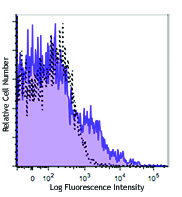
C57BL/6 mouse splenocytes stained with HMβ1-1 Alexa Fluor® 647 -

Rat frozen spleen section was fixed with 4% paraformaldehyde (PFA) for ten minutes at room temperature and blocked with 5% FBS for 30 minutes at room temperature. Then the section was stained with 10 µg/mL of CD3 (clone 1F4) Alexa Fluor® 488 (blue), 10 µg/mL of CD29 (clone HMβ1-1) Alexa Fluor® 647 (yellow), and 10 µg/mL of purified CD38 (clone 14.27) overnight at 4°C, followed by 5 µg/mL of anti-mouse IgG2b (clone RMG2b-1) Alexa Fluor® 594 (red) for two hours at room temperature. The image was captured by 10X objective.
| Cat # | Size | Price | Quantity Check Availability | Save | ||
|---|---|---|---|---|---|---|
| 102213 | 25 µg | 81€ | ||||
| 102214 | 100 µg | 184€ | ||||
CD29 is a 130 kD protein, also known as integrin β1, VLA-β chain, or GPIIa. It is a member of the integrin family, expressed broadly on leukocytes, endothelial cells, smooth muscle, and epithelial cells. In association with CD49a-f, CD29 forms the VLA-1 through VLA-6 complexes, respectively. It plays an important role in cell-cell or cell-matrix interaction. The HMß1-1 antibody reacts with both mouse and rat CD29. It is able to block cell adhesion and inhibit T cell proliferation.
Product DetailsProduct Details
- Verified Reactivity
- Mouse, Rat
- Antibody Type
- Monoclonal
- Host Species
- Armenian Hamster
- Immunogen
- Purified mouse VLA-4 (α4β1, CD49d/CD29)
- Formulation
- Phosphate-buffered solution, pH 7.2, containing 0.09% sodium azide.
- Preparation
- The antibody was purified by affinity chromatography and conjugated with Alexa Fluor® 647 under optimal conditions.
- Concentration
- 0.5 mg/ml
- Storage & Handling
- The antibody solution should be stored undiluted between 2°C and 8°C, and protected from prolonged exposure to light. Do not freeze.
- Application
-
FC - Quality tested
IHC-F - Verified - Recommended Usage
-
Each lot of this antibody is quality control tested by immunofluorescent staining with flow cytometric analysis. For flow cytometric staining, the suggested use of this reagent is ≤ 0.25 µg per 106 cells in 100 µl volume. For immunohistochemistry staining on frozen tissue sections, a concentration range of 2.5 - 10 µg/ml is suggested. It is recommended that the reagent be titrated for optimal performance for other applications.
* Alexa Fluor® 647 has a maximum emission of 668 nm when it is excited at 633nm / 635nm.
Alexa Fluor® and Pacific Blue™ are trademarks of Life Technologies Corporation.
View full statement regarding label licenses - Excitation Laser
-
Red Laser (633 nm)
- Application Notes
-
Additional reported applications (for the relevant formats) include: immunoprecipitation1, immunohistochemistry4 of acetone-fixed frozen sections, in vitro blocking of the adhesion of mouse tumor cell lines to extracellular matrix proteins and in vitro inhibition of T cell proliferative responses1, and in vivo inhibition of neutrophil migration2. The Ultra-LEAF™ purified antibody (Endotoxin < 0.01 EU/µg, Azide-Free, 0.2 µm filtered) is recommended for functional assays (Cat. No. 102235 and 102236).
-
Application References
(PubMed link indicates BioLegend citation) -
- Noto K, et al. 1995. Int. Immunol. 7:835.
- Ridger VC, et al. 2001. J. Immunol. 166:3484.
- Jia W, et al. 2005. Blood 106:3854.PubMed
- Economopoulou M, et al. 2005. Blood 106:3831.
- Lawson BR, et al. 2007. J. Immunol. 178:5366.
- Eisenmann KM, et al. 2007. J. Biol. Chem. doi:10.1074/jbc.M703243200.PubMed
- Hayashi Y, et al. 2008. Am J Physiol Gastrointest Liver Physiol. 294:G778. PubMed
- Kim DT, et al. 2008. Blood 111:2929.PubMed
- Hayashi Y, et al. 2008. J Pharmacol Exp Ther. 326:523. PubMed
- Carlson TR, et al. 2008. Development. 135:2193. PubMed
- Sangaletti S, et al. 2008. Cancer Res. 68:9050. (Block) PubMed
- Baker CM, et al. 2012. PNAS. PubMed.
- Hirokawa Y, et al. 2014. Am J Physiol Gastrointerest Liver Physiol. 306:547. PubMed
- Product Citations
-
- RRID
-
AB_492832 (BioLegend Cat. No. 102213)
AB_492831 (BioLegend Cat. No. 102214)
Antigen Details
- Structure
- Integrin family, 130 kD
- Distribution
-
Leukocytes, endothelial cells, smooth muscle, epithelial cells
- Function
- Adhesion
- Ligand/Receptor
- Extracellular matrix
- Cell Type
- Embryonic Stem Cells, Endothelial cells, Epithelial cells, Leukocytes, Mesenchymal Stem Cells, Tregs
- Biology Area
- Cell Adhesion, Cell Biology, Immunology, Innate Immunity, Stem Cells
- Molecular Family
- Adhesion Molecules, CD Molecules
- Antigen References
-
1. Noto K, et al. 1995. Int. Immunol. 7:835.
2. Springer TA. 1990. Nature 346:425. - Gene ID
- 16412 View all products for this Gene ID 24511 View all products for this Gene ID
- UniProt
- View information about CD29 on UniProt.org
Related Pages & Pathways
Pathways


Related FAQs
Other Formats
View All CD29 Reagents Request Custom Conjugation| Description | Clone | Applications |
|---|---|---|
| Biotin anti-mouse/rat CD29 | HMβ1-1 | FC |
| Purified anti-mouse/rat CD29 | HMβ1-1 | FC,IHC-F,IP,Block |
| FITC anti-mouse/rat CD29 | HMβ1-1 | FC |
| PE anti-mouse/rat CD29 | HMβ1-1 | FC |
| Alexa Fluor® 488 anti-mouse/rat CD29 | HMβ1-1 | FC,IHC-F |
| Alexa Fluor® 647 anti-mouse/rat CD29 | HMβ1-1 | FC,IHC-F |
| APC anti-mouse/rat CD29 | HMβ1-1 | FC |
| Alexa Fluor® 700 anti-mouse/rat CD29 | HMβ1-1 | FC |
| PE/Cyanine5 anti-mouse/rat CD29 | HMβ1-1 | FC |
| PE/Cyanine7 anti-mouse/rat CD29 | HMβ1-1 | FC |
| Pacific Blue™ anti-mouse/rat CD29 | HMβ1-1 | FC |
| APC/Cyanine7 anti-mouse/rat CD29 | HMβ1-1 | FC |
| PerCP/Cyanine5.5 anti-mouse/rat CD29 | HMβ1-1 | FC |
| PE/Dazzle™ 594 anti-mouse/rat CD29 | HMβ1-1 | FC |
| TotalSeq™-A0570 anti-mouse/rat CD29 | HMβ1-1 | PG |
| Ultra-LEAF™ Purified anti-mouse/rat CD29 | HMβ1-1 | FC,IHC-F,IP,Block |
| TotalSeq™-C0570 anti-mouse/rat CD29 Antibody | HMβ1-1 | PG |
| TotalSeq™-B0570 anti-mouse/rat CD29 | HMβ1-1 | PG |
Customers Also Purchased
Compare Data Across All Formats
This data display is provided for general comparisons between formats.
Your actual data may vary due to variations in samples, target cells, instruments and their settings, staining conditions, and other factors.
If you need assistance with selecting the best format contact our expert technical support team.
-
Biotin anti-mouse/rat CD29

Lou rat bone marrow cells stained with HMβ1-1 biotin, then d... 
C57BL/6 mouse splenocytes stained with HMβ1-1 PE -
Purified anti-mouse/rat CD29

Lou rat bone marrow cells stained with HMβ1-1 biotin, then d... 
C57BL/6 mouse splenocytes stained with HMβ1-1 PE. 
Rat frozen spleen section was fixed with 4% paraformaldehyde... -
FITC anti-mouse/rat CD29
Lou rat bone marrow cells stained with HMβ1-1 biotin, then d...
Lou rat bone marrow cells stained with HMβ1-1 biotin, then d... -
PE anti-mouse/rat CD29
Lou rat bone marrow cells stained with HMβ1-1 biotin, then d...
Lou rat bone marrow cells stained with HMβ1-1 biotin, then d... -
Alexa Fluor® 488 anti-mouse/rat CD29

C57BL/6 mouse splenocytes stained with HMβ1-1 Alexa Fluor® 4... 
C57BL/6 mouse frozen spleen section was fixed with 4% parafo... 
Rat frozen spleen section was fixed with 4% paraformaldehyde... -
Alexa Fluor® 647 anti-mouse/rat CD29

C57BL/6 mouse splenocytes stained with HMβ1-1 Alexa Fluor® 6... 
Rat frozen spleen section was fixed with 4% paraformaldehyde... -
APC anti-mouse/rat CD29

C57BL/6 mouse splenocytes stained with HMβ1-1 APC -
Alexa Fluor® 700 anti-mouse/rat CD29

C57BL/6 mouse bone marrow cells stained with HMβ1-1 Alexa Fl... -
PE/Cyanine5 anti-mouse/rat CD29

C57BL/6 mouse splenocytes stained with HMß1-1 PE/Cyanine5 -
PE/Cyanine7 anti-mouse/rat CD29

C57BL/6 mouse splenocytes stained with HMß1-1 PE/Cyanine7 -
Pacific Blue™ anti-mouse/rat CD29

C57BL/6 mouse bone marrow cells stained with HMβ1-1 Pacific ... -
APC/Cyanine7 anti-mouse/rat CD29

C57BL/6 bone marrow cells stained with HMb1-1 APC/Cyanine7 -
PerCP/Cyanine5.5 anti-mouse/rat CD29

C57BL/6 mouse bone marrow cells were stained with CD19 FITC ... -
PE/Dazzle™ 594 anti-mouse/rat CD29

C57BL/6 mouse bone marrow cells were stained with CD29 (clon... -
TotalSeq™-A0570 anti-mouse/rat CD29
-
Ultra-LEAF™ Purified anti-mouse/rat CD29
C57BL/6 mouse splenocytes stained with HMβ1-1 PE.
Lou rat bone marrow cells stained with HMβ1-1 biotin, then d... -
TotalSeq™-C0570 anti-mouse/rat CD29 Antibody
-
TotalSeq™-B0570 anti-mouse/rat CD29


Follow Us