A Sparkling Advancement for Flow Cytometry


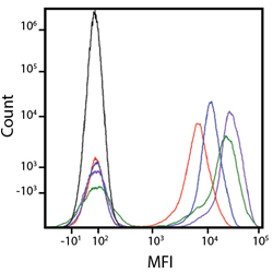
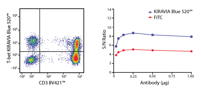
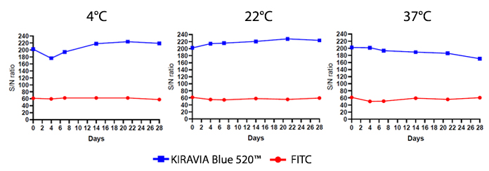
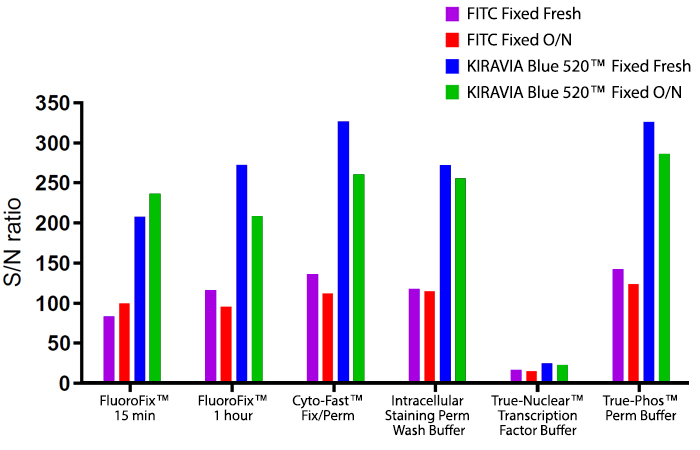
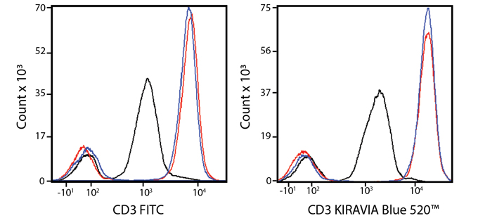
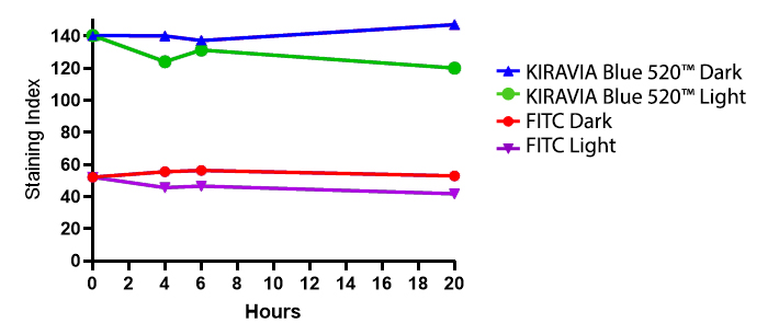
Introducing the KIRAVIA Dyes™, a brand new fluorescent chemistry to advance applications in multicolor flow cytometry and beyond. KIRAVIA is a coined term meaning "sparkling" in Japanese. KIRAVIA Blue 520™ is the first in a new family of fluorophores. This class of dyes employs a unique organic backbone that separates fluorophores to minimize quenching effects, thus allowing optimal and higher fluorophore to protein (F:P) ratios, surpassing what is possible with direct conjugations of single fluorophores like FITC.


View our full listing of KIRAVIA Blue 520™ antibodies and contact us to try a sample today.


KIRAVIA Dyes™ provided by Sony.


KIRAVIA Blue 520™ Spectra


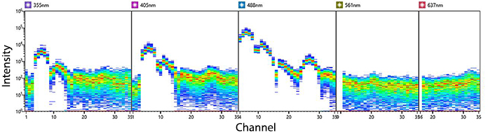
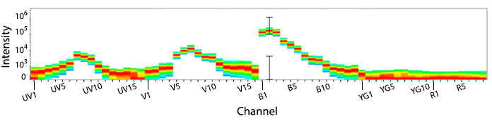
Emission spectra of KIRAVIA Blue 520™ as run on a (top) SONY ID7000™ Spectral Cell Analyzer or (bottom)  5-laser Cytek™ Aurora Spectral Cytometer.


A New Alternative to FITC With Minimal Spillover


In a multicolor panel, it is spectrally identical to FITC and only exhibits significant spillover into Spark Blue 550.


Spillover impact of KIRAVIA Blue 520™ into detection channels of a 5-laser Cytek™ Aurora Spectral Cytometer.


Amazing Resolution


KIRAVIA Blue 520™ is bright, on par with BD Horizon™ BB515 and, on average, is twice as bright as a FITC conjugate. KIRAVIA Blue 520™ can be used for cell surface, intracellular, and intranuclear antigens. It can be used to detect antigens with a variety of abundance levels, but it would ideally be saved for an antigen with ubiquitous and/or variable expression levels in the sample, specifically when used in complex multicolor panels (e.g. activation markers like CD25 and CD127).

Equivalent fluorophores comparison. Anti-human CD4 (clone SK3), conjugated to FITC (red), Alexa Fluor® 488 (blue), BD Horizon™ Brilliant Blue 515 (green), or KIRAVIA Blue 520™ (purple) was used to stain human lysed whole blood. This was compared to unstained cells (black).

Nuclear Staining and Titration (Human T-bet)


(Left) Anti-human CD3 (clone UCHT1) BV421™ and anti-human T-bet (clone 4B10) KIRAVIA Blue 520™ staining on human PBMCs. (Right) Titration curve with FITC or KIRAVIA Blue 520™ conjugated to anti-T-bet antibody.

Surface Staining (Human CCR7)

Human lysed whole blood was stained with anti-human CD3 APC (clone UCHT1) and anti-human CCR7 (clone G043H7) KIRAVIA Blue 520.

Intracellular Staining (Human IFN-γ)

PMA/ionomycin-stimulated (6 hours) human PBMCs stained with anti-human CD3 (clone UCHT1) APC and anti-IFN-γ (clone 4S.B3) KIRAVIA Blue 520™.


Stability Testing Data


Fluorophores can be exposed to a variety of conditions during the course of a flow cytometry experiment. In the data that follows, we provide useful information on how heat, light, and fixative can affect the performance of KIRAVIA Blue 520™.


Heat Stability


Anti-human CD3 (clone UCHT1) antibody conjugated to KIRAVIA Blue 520™ was incubated at the indicated temperatures over the course of 28 days. The antibodies were then used to stain human lysed whole blood cells. S/N ratio was determined as ratio of the MFI+/MFI- signals.


Fixative Stability


KIRAVIA Blue 520™ shows utility with most BioLegend fixation buffers. Similar to some fluorophores, KIRAVIA Blue 520™ can be sensitive to the nuclear permeabilization and fixation process if used as a surface stain conjugate. While a decrease in the MFI+ signal was observed when staining with the FITC and KIRAVIA Blue 520™ conjugates, the positive population could still be resolved easily. When building panels in these circumstances, the ability of a fluorophore conjugate to survive the fixation/permeabilization process will depend on the level of antigen expression and the sample type. For more panel building help and considerations, please contact our technical services group.


A guide to the fixatives used in this experiment:


Human PBMCs were stained with anti-human CD3 (clone UCHT1) conjugated to either FITC or KIRAVIA Blue 520™ and then fixed as indicated. (Top) Fixed Fresh samples were fixed according to their respective protocols and read on a cytometer immediately. O/N samples were fixed by the indicated buffer and stored overnight in Cyto-Last™ Buffer before being read on the cytometer. Signal to Noise (S/N) was determined as ratio of the MFI+/MFI- signals. (Bottom) Histogram overlay of the CD3 staining on PBMCs after treatment with True-Nuclear (black), Cyto-Fast™ (blue), or FluoroFix™ (red) as per their respective protocols.


Photostability


Human PBMCs were stained with anti-human CD3 (clone UCHT1) conjugated to either FITC or KIRAVIA Blue 520™. After staining, samples were fixed and stored in FluoroFix™ Buffer through the duration of the experiment. Samples were continually exposed to light or kept in the dark and read on a BD LSR Fortessa™ instrument.

ProductsHere

Login / Register
Remember me
Forgot your password? Reset password?
Create an Account