 Login / Register


- Clone
- FA-11 (See other available formats)
- Regulatory Status
- RUO
- Other Names
- Macrosialin
- Isotype
- Rat IgG2a
- Ave. Rating
- Submit a Review
- Product Citations
- publications

-

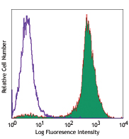
Thioglycolate-elicited BALB/c macrophages were fixed, permeabilized, and then stained with CD68 (clone FA-11) Alexa Fluor® 700 (filled histogram) or rat IgG2a, κ Alexa Fluor® 700 isotype control (open histogram).
| Cat # | Size | Price | Quantity Check Availability | Save | ||
|---|---|---|---|---|---|---|
| 137025 | 25 µg | £105 | ||||
| 137026 | 100 µg | £257 | ||||
Mouse CD68, also known as macrosialin, is an 85-115 kD member of the lysosomal-associated membrane protein (LAMP) family. It is a heavily glycosylated and predominantly intracellular protein, mainly in late endosomes. Macrosialin is the murine homolog to the human macrophage glycoprotein CD68. It is expressed on tissue macrophages, Langerhans cells and at low levels on dendritic cells. Lamp proteins may have functions relating to cell-cell interaction or cell-ligand interaction. The biological function of CD68 is not completely understood.
Product DetailsProduct Details
- Verified Reactivity
- Mouse
- Antibody Type
- Monoclonal
- Host Species
- Rat
- Immunogen
- Purified Con A receptor glycoproteins from the P815 cell line
- Formulation
- Phosphate-buffered solution, pH 7.2, containing 0.09% sodium azide.
- Preparation
- The antibody was purified by affinity chromatography and conjugated with Alexa Fluor® 700 under optimal conditions.
- Concentration
- 0.5 mg/ml
- Storage & Handling
- The antibody solution should be stored undiluted between 2°C and 8°C, and protected from prolonged exposure to light. Do not freeze.
- Application
-
ICFC - Quality tested
- Recommended Usage
-
Each lot of this antibody is quality control tested by intracellular immunofluorescent staining with flow cytometric analysis. For flow cytometric staining, the suggested use of this reagent is ≤ 0.25 µg per million cells in 100 µl volume. It is recommended that the reagent be titrated for optimal performance for each application.
* Alexa Fluor® 700 has a maximum emission of 719 nm when it is excited at 633 nm / 635 nm. Prior to using Alexa Fluor® 700 conjugate for flow cytometric analysis, please verify your flow cytometer's capability of exciting and detecting the fluorochrome.
Alexa Fluor® and Pacific Blue™ are trademarks of Life Technologies Corporation.
View full statement regarding label licenses - Excitation Laser
-
Red Laser (633 nm)
- Application Notes
-
Additional reported (for relevant formats) applications include: immunoprecipitation1, 2, Western Blot1, 2, immunohistochemical staining of frozen sections2 and paraformaldehyde-fixed paraffin-embedded sections3, and spatial biology (IBEX)9,10.
-
Application References
(PubMed link indicates BioLegend citation) -
- Silva RP, et al. 1999. Biochem. J. 338:687. (IP, WB)
- Rabinowitz SS, et al. 1991. J. Exp. Med. 174:827. (IP, WB, IHC)
- Wu J, et al. 2008. P. Natl. Acad. Sci. USA 105:16934. (IHC)
- Kayama H, et al. 2012. PNAS. 109:5010. PubMed
- Park S, et al. 2013. Biomaterials. 34:598. PubMed
- Guiducci C, et al. 2013. J Exp Med. 210:2903. PubMed
- McKinstry SU, et al. 2014. J Neurosci. 34:9455. PubMed
- Li X, et al. 2015. J Am Heart Assoc. 6:4. PubMed
- Radtke AJ, et al. 2020. Proc Natl Acad Sci U S A. 117:33455-65. (SB) PubMed
- Radtke AJ, et al. 2022. Nat Protoc. 17:378-401. (SB) PubMed
- RRID
-
AB_2783096 (BioLegend Cat. No. 137025)
AB_2783097 (BioLegend Cat. No. 137026)
Antigen Details
- Structure
- A member of the lysosomal-associated membrane protein (lamp) family.
- Distribution
-
Expressed on tissue macrophages, Langerhans cells, and at low levels on dendritic cells.
- Function
- Involved in cell-cell interaction or cell-ligand interaction, still not completely understood.
- Cell Type
- Antigen-presenting cells, Dendritic cells, Langerhans cells, Leukocytes, Macrophages
- Biology Area
- Cell Biology, Immunology, Innate Immunity, Neuroscience, Neuroscience Cell Markers
- Molecular Family
- Adhesion Molecules, CD Molecules, Innate Immune Signaling
- Antigen References
-
1. Ramprasad MP, et al. 1996. Proc. Natl. Acad. Sci. USA 93:14833.
2. Smith MJ, et al. 1987. J. Cell. Sci. 87:113. - Gene ID
- 12514 View all products for this Gene ID
- UniProt
- View information about CD68 on UniProt.org
Related Pages & Pathways
Pathways


Related FAQs
Other Formats
View All CD68 Reagents Request Custom Conjugation| Description | Clone | Applications |
|---|---|---|
| Purified anti-mouse CD68 | FA-11 | ICFC,FC,IP,WB,IHC-F |
| Alexa Fluor® 647 anti-mouse CD68 | FA-11 | ICFC,FC,SB |
| FITC anti-mouse CD68 | FA-11 | ICFC,FC |
| APC anti-mouse CD68 | FA-11 | ICFC,FC |
| PerCP/Cyanine5.5 anti-mouse CD68 | FA-11 | ICFC,FC |
| Alexa Fluor® 488 anti-mouse CD68 | FA-11 | ICFC,FC,3D IHC |
| PE anti-mouse CD68 | FA-11 | ICFC,FC |
| PE/Cyanine7 anti-mouse CD68 | FA-11 | ICFC,FC |
| Brilliant Violet 421™ anti-mouse CD68 | FA-11 | ICFC,FC,SB |
| Alexa Fluor® 594 anti-mouse CD68 | FA-11 | IHC-F,3D IHC |
| APC/Cyanine7 anti-mouse CD68 | FA-11 | ICFC,FC |
| Brilliant Violet 605™ anti-mouse CD68 | FA-11 | ICFC,FC |
| Brilliant Violet 711™ anti-mouse CD68 | FA-11 | ICFC |
| Alexa Fluor® 700 anti-mouse CD68 | FA-11 | ICFC |
| Pacific Blue™ anti-mouse CD68 | FA-11 | ICFC |
| TotalSeq™-A0560 anti-mouse CD68 | FA-11 | PG |
| TotalSeq™-C0560 anti-mouse CD68 | FA-11 | PG |
| Brilliant Violet 785™ anti-mouse CD68 | FA-11 | ICFC |
| TotalSeq™-B0560 anti-mouse CD68 | FA-11 | PG |
| Spark YG™ 570 anti-mouse CD68 | FA-11 | IHC-F |
| APC/Fire™ 750 anti-mouse CD68 | FA-11 | ICFC |
Customers Also Purchased
Compare Data Across All Formats
This data display is provided for general comparisons between formats.
Your actual data may vary due to variations in samples, target cells, instruments and their settings, staining conditions, and other factors.
If you need assistance with selecting the best format contact our expert technical support team.
-
Purified anti-mouse CD68

Thioglycolate-elicited Balb/c peritoneal macrophages intrace... -
Alexa Fluor® 647 anti-mouse CD68
Thioglycolate-elicited Balb/c peritoneal macrophages intrace... -
FITC anti-mouse CD68

Thioglycolate-elicited Balb/c peritoneal macrophages intrace... -
APC anti-mouse CD68

Thioglycolate-elicited Balb/c peritoneal macrophages intrace... -
PerCP/Cyanine5.5 anti-mouse CD68

Thioglycolate-elicited BALB/c macrophages were fixed, permea... -
Alexa Fluor® 488 anti-mouse CD68

Thioglycolate-elicited Balb/c peritoneal macrophages intrace... 
Paraformaldehyde-fixed (4%), 500 μm-thick mouse spleen secti... -
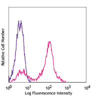
PE anti-mouse CD68

Thioglycolate-elicited BALB/c peritoneal macrophages intrace... -
PE/Cyanine7 anti-mouse CD68

Thioglycolate-elicited BALB/c macrophages were fixed, permea... -
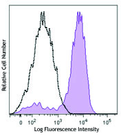
Brilliant Violet 421™ anti-mouse CD68

Thioglycolate-elicited BALB/c macrophages were fixed, permea... 
Confocal image of C57BL/6 mouse spleen sample acquired using... 
Confocal image of C57BL/6 mouse thymus sample acquired using... 
Confocal image of C57BL/6 mouse lung sample acquired using t... 
Mice were injected subcutaneously with sheep red blood cells... -
Alexa Fluor® 594 anti-mouse CD68

C57BL/6 mouse frozen spleen section was fixed with 4% parafo... 
Paraformaldehyde-fixed (4%), 500 µm-thick mouse spleen secti... -
APC/Cyanine7 anti-mouse CD68

Thioglycolate-elicited BALB/c macrophages were fixed, permea... -
Brilliant Violet 605™ anti-mouse CD68

Thioglycolate-elicited BALB/c macrophages were fixed, permea... -
Brilliant Violet 711™ anti-mouse CD68

Thioglycolate-elicited BALB/c macrophages were fixed, permea... -
Alexa Fluor® 700 anti-mouse CD68

Thioglycolate-elicited BALB/c macrophages were fixed, permea... -
Pacific Blue™ anti-mouse CD68

Thioglycolate-elicited BALB/c macrophages were fixed, permea... -
TotalSeq™-A0560 anti-mouse CD68
-
TotalSeq™-C0560 anti-mouse CD68
-
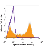
Brilliant Violet 785™ anti-mouse CD68

Thioglycolate-elicited Balb/c peritoneal macrophages were su... -
TotalSeq™-B0560 anti-mouse CD68
-
Spark YG™ 570 anti-mouse CD68

C57BL/6 mouse frozen spleen section was fixed with 4% parafo... -
APC/Fire™ 750 anti-mouse CD68

Thioglycollate-elicited BALB/c macrophages were fixed, perm...


Follow Us