


- Clone
- 3D6.112 (See other available formats)
- Regulatory Status
- RUO
- Other Names
- Sialic acid binding Ig-like lectin 1 (Siglec-1), Sialoadhesin (Sn)
- Isotype
- Rat IgG2a, κ
- Ave. Rating
- Submit a Review
- Product Citations
- publications

-

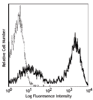
C57BL/6 splenocytes were stained with F4/80 APC, Ly-6G PerCP, and CD169 (clone 3D6.112) PE (top) or rat IgG2a, κ PE isotype control (bottom). Data was analyzed by gating on Ly-6G-negative cell population. -

-

Fresh, frozen mouse spleen was stained with purified CD169 clone 3D6.112 conjugated and detected with a Cy3 CODEX™ oligonucleotide duplex (red). Samples were counterstained with B220 A647 (green). Data generated at Akoya Biosciences, Inc. using the CODEX™ technology.
| Cat # | Size | Price | Save |
|---|---|---|---|
| 142401 | 25 µg | ¥24,640 | |
| 142402 | 100 µg | ¥50,380 |
CD169, also known as Siglec-1 and Sialoadhesin (Sn), is a type I lectin containing 17 immunoglobulin (Ig) domains (one variable domain and 16 constant domains). CD169 binds to sialic acids, which can be found on PSGL-1, CD43, CD206, and CD227. By its affinity to α2, 3-linked sialic acid, it is involved in macrophage binding to different cell types such as granulocytes, monocytes, NK, B, and T cells. CD169 was initially identified as a sialic acid-dependent sheep erythrocyte receptor (SER) on resident bone marrow cells of mice. It has been identified as highly expressed on resident bone marrow macrophages which plays an important role in retention of stem cells in mesenchymal stem cell niche. It is also found on some specific subsets of tissue macrophages in spleen, lymph nodes, bone marrow, liver, colon, lungs, and cancer cells. Evidence suggest that CD169-positive macrophages serve as lymph node-resident APCs to dominate early activation of tumor antigen-specific CD8+ T cells and invariant NK cell.
Product DetailsProduct Details
- Verified Reactivity
- Mouse
- Antibody Type
- Monoclonal
- Host Species
- Rat
- Immunogen
- Purified Native Sialoadhesin from spleen
- Formulation
- Phosphate-buffered solution, pH 7.2, containing 0.09% sodium azide.
- Preparation
- The antibody was purified by affinity chromatography.
- Concentration
- 0.5 mg/ml
- Storage & Handling
- The antibody solution should be stored undiluted between 2°C and 8°C.
- Application
-
FC - Quality tested
IHC-F - Verified
SB - Community verified - Recommended Usage
-
Each lot of this antibody is quality control tested by immunofluorescent staining with flow cytometric analysis. For flow cytometric staining, the suggested use of this reagent is ≤0.5 µg per million cells in 100 µl volume. It is recommended that the reagent be titrated for optimal performance for each application.
- Application Notes
-
Additional reported applications (for the relevant formats) include: immunohistochemical staining in frozen tissue sections1, and spatial biology (IBEX)4,5.
- Additional Product Notes
-
This product has been verified for IHC-F (Immunohistochemistry - frozen tissue sections) on the NanoString GeoMx® Digital Spatial Profiler. The GeoMx® enables researchers to perform spatial analysis of protein and RNA targets in FFPE and fresh frozen human and mouse samples. For more information about our spatial biology products and the GeoMx® platform, please visit our spatial biology page.
-
Application References
(PubMed link indicates BioLegend citation) - Product Citations
-
- RRID
-
AB_10915134 (BioLegend Cat. No. 142401)
AB_10916523 (BioLegend Cat. No. 142402)
Antigen Details
- Structure
- Type I single membrane-spanning lectin containing 17 immunoglobulin (Ig) domains, belongs to the immunoglobulin superfamily.
- Distribution
-
Macrophages in spleen, lymph nodes, bone marrow, liver, colon and lungs.
- Function
- Adhesion.
- Ligand/Receptor
- PSGL-1, CD43, CD206 and CD227.
- Cell Type
- Macrophages
- Biology Area
- Cell Biology, Immunology
- Molecular Family
- Adhesion Molecules, CD Molecules, Protein Kinases/Phosphatase, Siglec Molecules
- Antigen References
-
1. Chow A, et al. 2011. J. Exp. Med. 208:261.
2. Asano K, et al. 2011. Immunity 34:85.
3. Xiong YS, et al. 2009. Clin. Biochem. 42:1057.
4. Varki A, et al. 2009. Glycoconj. J. 26:231.
5. Rempel H, et al. 2008. PLoS One 3:e1967.
6. Crocker PR, et al. 2001. Trends Immunol. 22:337.
7. Hartnell A, et al. 2001. Blood 97:288.
8. Crocker PR, et al. 1985. J. Exp. Med. 162:993. - Gene ID
- 20612 View all products for this Gene ID
- UniProt
- View information about CD169 on UniProt.org


Related FAQs
- If an antibody clone has been previously successfully used in IBEX in one fluorescent format, will other antibody formats work as well?
-
It’s likely that other fluorophore conjugates to the same antibody clone will also be compatible with IBEX using the same sample fixation procedure. Ultimately a directly conjugated antibody’s utility in fluorescent imaging and IBEX may be specific to the sample and microscope being used in the experiment. Some antibody clone conjugates may perform better than others due to performance differences in non-specific binding, fluorophore brightness, and other biochemical properties unique to that conjugate.
- Will antibodies my lab is already using for fluorescent or chromogenic IHC work in IBEX?
-
Fundamentally, IBEX as a technique that works much in the same way as single antibody panels or single marker IF/IHC. If you’re already successfully using an antibody clone on a sample of interest, it is likely that clone will have utility in IBEX. It is expected some optimization and testing of different antibody fluorophore conjugates will be required to find a suitable format; however, legacy microscopy techniques like chromogenic IHC on fixed or frozen tissue is an excellent place to start looking for useful antibodies.
- Are other fluorophores compatible with IBEX?
-
Over 18 fluorescent formats have been screened for use in IBEX, however, it is likely that other fluorophores are able to be rapidly bleached in IBEX. If a fluorophore format is already suitable for your imaging platform it can be tested for compatibility in IBEX.
- The same antibody works in one tissue type but not another. What is happening?
-
Differences in tissue properties may impact both the ability of an antibody to bind its target specifically and impact the ability of a specific fluorophore conjugate to overcome the background fluorescent signal in a given tissue. Secondary stains, as well as testing multiple fluorescent conjugates of the same clone, may help to troubleshoot challenging targets or tissues. Using a reference control tissue may also give confidence in the specificity of your staining.
- How can I be sure the staining I’m seeing in my tissue is real?
-
In general, best practices for validating an antibody in traditional chromogenic or fluorescent IHC are applicable to IBEX. Please reference the Nature Methods review on antibody based multiplexed imaging for resources on validating antibodies for IBEX.
Other Formats
View All CD169 Reagents Request Custom Conjugation| Description | Clone | Applications |
|---|---|---|
| Purified anti-mouse CD169 (Siglec-1) | 3D6.112 | FC,IHC-F,SB |
| PE anti-mouse CD169 (Siglec-1) | 3D6.112 | FC,SB |
| FITC anti-mouse CD169 (Siglec-1) | 3D6.112 | FC |
| Alexa Fluor® 647 anti-mouse CD169 (Siglec-1) | 3D6.112 | FC,IHC-F,3D IHC |
| PerCP/Cyanine5.5 anti-mouse CD169 (Siglec-1) | 3D6.112 | FC |
| PE/Cyanine7 anti-mouse CD169 (Siglec-1) | 3D6.112 | FC |
| Brilliant Violet 605™ anti-mouse CD169 (Siglec-1) | 3D6.112 | FC |
| APC anti-mouse CD169 (Siglec-1) | 3D6.112 | FC |
| Alexa Fluor® 594 anti-mouse CD169 (Siglec-1) | 3D6.112 | IHC-F,3D IHC |
| Alexa Fluor® 488 anti-mouse CD169 (Siglec-1) | 3D6.112 | IHC-F,3D IHC |
| Brilliant Violet 421™ anti-mouse CD169 (Siglec-1) | 3D6.112 | IHC-F |
| PE/Dazzle™ 594 anti-mouse CD169 (Siglec-1) | 3D6.112 | FC |
| TotalSeq™-A0440 anti-mouse CD169 (Siglec-1) | 3D6.112 | PG |
| TotalSeq™-C0440 anti-mouse CD169 (Siglec-1) | 3D6.112 | PG |
| TotalSeq™-B0440 anti-mouse CD169 (Siglec-1) | 3D6.112 | PG |
| Biotin anti-mouse CD169 (Siglec-1) | 3D6.112 | FC |
Customers Also Purchased
Compare Data Across All Formats
This data display is provided for general comparisons between formats.
Your actual data may vary due to variations in samples, target cells, instruments and their settings, staining conditions, and other factors.
If you need assistance with selecting the best format contact our expert technical support team.
-
Purified anti-mouse CD169 (Siglec-1)

C57BL/6 splenocytes were stained with F4/80 APC, Ly-6G PerCP... 

Fresh, frozen mouse spleen was stained with purified CD169 c... -
PE anti-mouse CD169 (Siglec-1)
C57BL/6 splenocytes were stained with F4/80 APC, Ly-6G PerCP...

C57BL/6 inguinal lymph node labeled in-vivo with PE anti-mou... 
Live intravital mouse spleen imaging. PE CD169 (red) (clone ... 
Fixed whole mount mouse spleen imaging sectioned after intra... 
Confocal image of C57BL/6 mouse spleen sample acquired using... -
FITC anti-mouse CD169 (Siglec-1)

C57BL/6 mouse bone marrow cells were stained with F4/80 PE, ... 
-
Alexa Fluor® 647 anti-mouse CD169 (Siglec-1)

C57BL/6 mouse bone marrow cells were stained with F4/80 PE, ... 

C57BL/6 mouse frozen lymph node section was fixed with 4% pa... Formalin-fixed, 300 micron-thick mouse spleen section was bl... 
Paraformaldehyde-fixed (1%), 500 μm-thick mouse spleen secti... -
PerCP/Cyanine5.5 anti-mouse CD169 (Siglec-1)

C57BL/6 mouse bone marrow cells were stained with Ly-6G APC ... -
PE/Cyanine7 anti-mouse CD169 (Siglec-1)

C57BL/6 mouse bone marrow cells were stained with Ly-6G FITC... -
Brilliant Violet 605™ anti-mouse CD169 (Siglec-1)

C57BL/6 mouse bone marrow cells were stained with Ly-6G FITC... 
-
APC anti-mouse CD169 (Siglec-1)

C57BL/6 mouse bone marrow cells were stained with Ly-6G PE a... 
-
Alexa Fluor® 594 anti-mouse CD169 (Siglec-1)

C57BL/6 mouse frozen spleen section was fixed with 4% parafo... 
Paraformaldehyde-fixed (4%), 500 µm-thick mouse spleen secti... -
Alexa Fluor® 488 anti-mouse CD169 (Siglec-1)

C57BL/6 mouse frozen spleen section was fixed with 4% parafo... 
Dissected C57/B6 mouse spleen was immersed in 4% paraformal... 
Formalin-fixed, 400 micron-thick mouse spleen section was bl... -
Brilliant Violet 421™ anti-mouse CD169 (Siglec-1)

C57BL/6 mouse frozen spleen section was fixed with 4% parafo... -
PE/Dazzle™ 594 anti-mouse CD169 (Siglec-1)

C57BL/6 mouse bone marrow cells were stained with Ly-6G FITC... -
TotalSeq™-A0440 anti-mouse CD169 (Siglec-1)
-
TotalSeq™-C0440 anti-mouse CD169 (Siglec-1)
-
TotalSeq™-B0440 anti-mouse CD169 (Siglec-1)
-
Biotin anti-mouse CD169 (Siglec-1)

C57BL/6 mouse bone marrow cells were stained with anti-mouse...


Follow Us