 Login/Register


- Clone
- IM7 (See other available formats)
- Regulatory Status
- RUO
- Other Names
- Hermes, Pgp-1, H-CAM, HUTCH-1, ECMR III, gp85, Ly-24
- Isotype
- Rat IgG2b, κ
- Ave. Rating
- Submit a Review
- Product Citations
- publications

-

C57BL/6 mouse splenocytes stained with IM7 Alexa Fluor® 488 -

Confocal image of human lymph node sample acquired using the IBEX method of highly multiplexed antibody-based imaging: CD21 (yellow) in Cycle 3 and CD44 (blue) in Cycle 6. Tissues were prepared using ~1% (vol/vol) formaldehyde and a detergent. Following fixation, samples are immersed in 30% (wt/vol) sucrose for cryoprotection. Images are courtesy of Drs. Andrea J. Radtke and Ronald N. Germain of the Center for Advanced Tissue Imaging (CAT-I) in the National Institute of Allergy and Infectious Diseases (NIAID, NIH). -

Confocal image of human jejunum sample acquired using the IBEX method of highly multiplexed antibody-based imaging: EpCAM (blue) in Cycle 1 and CD44 (yellow) in Cycle 6. Tissues were prepared using ~1% (vol/vol) formaldehyde and a detergent. Following fixation, samples are immersed in 30% (wt/vol) sucrose for cryoprotection. Images are courtesy of Drs. Andrea J. Radtke and Ronald N. Germain of the Center for Advanced Tissue Imaging (CAT-I) in the National Institute of Allergy and Infectious Diseases (NIAID, NIH). -

Confocal image of human skin sample acquired using the IBEX method of highly multiplexed antibody-based imaging: Keratin 14 (yellow) in Cycle 4 and CD44 (purple) in Cycle 4. Tissues were prepared using ~1% (vol/vol) formaldehyde and a detergent. Following fixation, samples are immersed in 30% (wt/vol) sucrose for cryoprotection. Images are courtesy of Drs. Andrea J. Radtke and Ronald N. Germain of the Center for Advanced Tissue Imaging (CAT-I) in the National Institute of Allergy and Infectious Diseases (NIAID, NIH).
| Cat # | Size | Price | Quantity Check Availability | Save | ||
|---|---|---|---|---|---|---|
| 103015 | 25 µg | $101 | ||||
| 103016 | 100 µg | $229 | ||||
CD44 is a 80-95 kD glycoprotein also known as Hermes, Pgp1, H-CAM, or HUTCH. It is expressed on all leukocytes, endothelial cells, hepatocytes, and mesenchymal cells. As B and T cells become activated or progress to the memory stage, CD44 expression increases from low or mid levels to high levels. Thus, CD44 has been reported to be a valuable marker for memory cell subsets. High CD44 expression on Treg cells has been associated with potent suppressive function via high production of IL-10. CD44 is an adhesion molecule involved in leukocyte attachment to and rolling on endothelial cells, homing to peripheral lymphoid organs and to the sites of inflammation, and leukocyte aggregation.
Product DetailsProduct Details
- Verified Reactivity
- Mouse, Human
- Reported Reactivity
- Chimpanzee, Baboon, Cynomolgus, Rhesus, Squirrel Monkey, Horse, Cow, Pig, Dog, Cat
- Antibody Type
- Monoclonal
- Host Species
- Rat
- Immunogen
- Dexamethasone-induced myeloid leukemia M1 cells
- Formulation
- Phosphate-buffered solution, pH 7.2, containing 0.09% sodium azide.
- Preparation
- The antibody was purified by affinity chromatography and conjugated with Alexa Fluor® 488 under optimal conditions.
- Concentration
- 0.5 mg/ml
- Storage & Handling
- The antibody solution should be stored undiluted between 2°C and 8°C, and protected from prolonged exposure to light. Do not freeze.
- Application
-
FC - Quality tested
SB - Reported in the literature, not verified in house - Recommended Usage
-
Each lot of this antibody is quality control tested by immunofluorescent staining with flow cytometric analysis. For flow cytometric staining, the suggested use of this reagent is ≤1.0 µg per million cells in 100 µl volume. It is recommended that the reagent be titrated for optimal performance for each application.
* Alexa Fluor® 488 has a maximum emission of 519 nm when it is excited at 488 nm.
Alexa Fluor® and Pacific Blue™ are trademarks of Life Technologies Corporation.
View full statement regarding label licenses - Excitation Laser
-
Blue Laser (488 nm)
- Application Notes
-
Clone IM7 has been reported to recognize an epitope common to alloantigens and all isoforms of CD4417,18 that is located between amino acids 145 and 18620. This clone has been verified for immunocytochemistry (ICC) and frozen immunohistochemistry (IHC-F). Additional reported applications (for the relevant formats) include: immunohistochemistry of acetone-fixed frozen sections and formalin-fixed paraffin-embedded sections6,7, complement-mediated cytotoxicity1, immunoprecipitation1,3, in vivo inhibition of DTH4,5, and spatial biology (IBEX)23,24. The Ultra-LEAF™ purified antibody (Endotoxin < 0.01 EU/µg, Azide-Free, 0.2 µm filtered) is recommended for functional assays (Cat. No. 103046, 103065 - 103069).
Cross-reactivity to ferret has been reported by a collaborator, but not verified in house. - Additional Product Notes
-
Iterative Bleaching Extended multi-pleXity (IBEX) is a fluorescent imaging technique capable of highly-multiplexed spatial analysis. The method relies on cyclical bleaching of panels of fluorescent antibodies in order to image and analyze many markers over multiple cycles of staining, imaging, and, bleaching. It is a community-developed open-access method developed by the Center for Advanced Tissue Imaging (CAT-I) in the National Institute of Allergy and Infectious Diseases (NIAID, NIH).
-
Application References
(PubMed link indicates BioLegend citation) -
- Trowbridge IS, et al. 1982. Immunogenetics 15:299. (ICFC, IP, CMCD)
- Katoh S, et al. 1994. J. Immunol. 153:3440. (ELISA)
- Budd RC, et al. 1987. J. Immunol. 138:3120. (IP)
- Camp RL, et al. 1993. J. Exp. Med. 178:497. (Block)
- Weiss JM, et al. 1997. J. Cell Biol. 137:1137. (Block)
- Frank NY, et al. 2005. Cancer Res. 65:4320. (IHC) PubMed
- Cuff CA, et al. 2001. J. Clin. Invest. 108:1031. (IHC)
- Lee JW, et al. 2006. Nature Immunol. 8:181.
- Zhang N, et al. 2005. J. Immunol. 174:6967. PubMed
- Huabiao C, et al. 2005. J. Immunol. 175:591. PubMed
- Gui J, et al. 2007. Int. Immunol. 19:1201. PubMed
- Wang XY, et al. 2008. Blood 111:2436. PubMed
- Kenna TJ, et al. 2008. Blood 111:2091. PubMed
- Yamazaki J, et al. 2009. Blood PubMed
- Kmieciak M, et al. 2009. J. Transl. Med. 7:89. (FC) PubMed
- Chen YW, et al. 2010. Mol. Cancer Ther. 9:2879. PubMed
- Zheng Z, et al. 1995. J. Cell. Biol. 130:485.
- Wiranowska M, et al. 2010. Int. J. Cancer 127:532.
- Hirokawa Y, et al. 2014. Am J Physiol Gastrointerest Liver Physiol. 306:547. PubMed
- Sandmaier BM, et al. 1998. Blood 91:3494.
- Yang Y, et al. 2015. Hypertension. 65:1047. PubMed
- Peterson VM, et al. 2017. Nat. Biotechnol. 35:936. (PG)
- Radtke AJ, et al. 2020. Proc Natl Acad Sci U S A. 117:33455-65. (SB) PubMed
- Radtke AJ, et al. 2022. Nat Protoc. 17:378-401. (SB) PubMed
- Product Citations
-
- RRID
-
AB_493678 (BioLegend Cat. No. 103015)
AB_493679 (BioLegend Cat. No. 103016)
Antigen Details
- Structure
- Variable splicing of CD44 gene generates many CD44 isoforms, 80-95 kD
- Distribution
-
All leukocytes, epithelial cells, endothelial cells, hepatocytes, mesenchymal cells
- Function
- Leukocyte attachment and rolling on endothelial cells, stromal cells and ECM
- Ligand/Receptor
- Hyaluronan, MIP-1β, fibronectin, collagen
- Cell Type
- B cells, Endothelial cells, Epithelial cells, Leukocytes, Mesenchymal cells, Mesenchymal Stem Cells, Tregs
- Biology Area
- Cell Adhesion, Cell Biology, Immunology, Stem Cells
- Molecular Family
- Adhesion Molecules, CD Molecules
- Antigen References
-
1. Barclay AN, et al. 1997. The Leukocyte Antigen FactsBook Academic Press.
2. Haynes BF, et al. 1991. Cancer Cells 3:347.
3. Goldstein LA, et al. 1989. Cell 56:1063.
4. Mikecz K, et al. 1995. Nat. Med. 1:558.
5. Hegde V, et al. 2008. J. Leukocyte Biol. 84:134.
6. Liu T, et al. 2009. Biol. Direct 4:40. - Gene ID
- 12505 View all products for this Gene ID 960 View all products for this Gene ID
- UniProt
- View information about CD44 on UniProt.org
Related Pages & Pathways
Pathways


Related FAQs
- If an antibody clone has been previously successfully used in IBEX in one fluorescent format, will other antibody formats work as well?
-
It’s likely that other fluorophore conjugates to the same antibody clone will also be compatible with IBEX using the same sample fixation procedure. Ultimately a directly conjugated antibody’s utility in fluorescent imaging and IBEX may be specific to the sample and microscope being used in the experiment. Some antibody clone conjugates may perform better than others due to performance differences in non-specific binding, fluorophore brightness, and other biochemical properties unique to that conjugate.
- Will antibodies my lab is already using for fluorescent or chromogenic IHC work in IBEX?
-
Fundamentally, IBEX as a technique that works much in the same way as single antibody panels or single marker IF/IHC. If you’re already successfully using an antibody clone on a sample of interest, it is likely that clone will have utility in IBEX. It is expected some optimization and testing of different antibody fluorophore conjugates will be required to find a suitable format; however, legacy microscopy techniques like chromogenic IHC on fixed or frozen tissue is an excellent place to start looking for useful antibodies.
- Are other fluorophores compatible with IBEX?
-
Over 18 fluorescent formats have been screened for use in IBEX, however, it is likely that other fluorophores are able to be rapidly bleached in IBEX. If a fluorophore format is already suitable for your imaging platform it can be tested for compatibility in IBEX.
- The same antibody works in one tissue type but not another. What is happening?
-
Differences in tissue properties may impact both the ability of an antibody to bind its target specifically and impact the ability of a specific fluorophore conjugate to overcome the background fluorescent signal in a given tissue. Secondary stains, as well as testing multiple fluorescent conjugates of the same clone, may help to troubleshoot challenging targets or tissues. Using a reference control tissue may also give confidence in the specificity of your staining.
- How can I be sure the staining I’m seeing in my tissue is real?
-
In general, best practices for validating an antibody in traditional chromogenic or fluorescent IHC are applicable to IBEX. Please reference the Nature Methods review on antibody based multiplexed imaging for resources on validating antibodies for IBEX.
Other Formats
View All CD44 Reagents Request Custom ConjugationCustomers Also Purchased
Compare Data Across All Formats
This data display is provided for general comparisons between formats.
Your actual data may vary due to variations in samples, target cells, instruments and their settings, staining conditions, and other factors.
If you need assistance with selecting the best format contact our expert technical support team.
-
APC anti-mouse/human CD44

C57BL/6 mouse splenocytes stained with IM7 APC -
Biotin anti-mouse/human CD44

C57BL/6 mouse splenocytes were stained with biotinylated CD4... 
Mouse embryonic fibroblast NIH/3T3 cell line was fixed with ... -
FITC anti-mouse/human CD44

C57BL/6 splenocytes stained with IM7 FITC 
Human peripheral blood lymphocytes stained with IM7 FITC -
PE/Cyanine5 anti-mouse/human CD44

C57BL/6 mouse splenocytes stained with IM7 PE/Cyanine5 -
Purified anti-mouse/human CD44

C57BL/6 mouse splenocytes stained with purified IM7, followe... 
Fresh, frozen mouse spleen was stained with purified CD44 cl... -
Brilliant Violet 605™ anti-mouse/human CD44

C57BL/6 mouse splenocytes were stained with CD44 (clone IM7)... -
PE anti-mouse/human CD44

C57BL/6 mouse splenocytes stained with IM7 PE 
Human peripheral blood lymphocytes stained with IM7 PE -
Alexa Fluor® 488 anti-mouse/human CD44

C57BL/6 mouse splenocytes stained with IM7 Alexa Fluor® 488 
Confocal image of human lymph node sample acquired using the... 
Confocal image of human jejunum sample acquired using the IB... 
Confocal image of human skin sample acquired using the IBEX ... -
Alexa Fluor® 647 anti-mouse/human CD44
IM7_Alx647_091907")
C57BL/6 mouse splenocytes stained with IM7 Alexa Fluor® 647 IM7_AF647_CD44_Antibody_IF_092013")
MDA-MB231 breast cancer cell line was stained with 5 µg/mL a... 46_Mouse_Lung_Btub3_CD68_CD44")
Confocal image of C57BL/6 mouse lung sample acquired using t... 65_Mouse_Lymph_Node_CD44")
Mice were injected subcutaneously with sheep red blood cells... 14_Human_Spleen_CD49a_CD44")
Confocal image of human spleen sample acquired using the IBE... 19_Human_Liver_Keratin7_CD44_CD45")
Confocal image of human liver sample acquired using the IBEX... -
Pacific Blue™ anti-mouse/human CD44

C57BL/6 Mouse Splenocytes stained with IM7 Pacific Blue&trad... -
Alexa Fluor® 700 anti-mouse/human CD44

C57BL/6 mouse splenocytes stained with IM7 Alexa Fluor® 700 
Confocal image of C57BL/6 mouse liver sample acquired using ... -
PE/Cyanine7 anti-mouse/human CD44

C57BL/6 mouse splenocytes stained with IM7 PE/Cyanine7 -
APC/Cyanine7 anti-mouse/human CD44

C57BL/6 splenocytes stained with IM7 APC/Cyanine7 -
PerCP/Cyanine5.5 anti-mouse/human CD44

C57BL/6 mouse splenocytes were stained with CD44 (clone IM7)... -
PerCP anti-mouse/human CD44

C57BL/6 splenocytes stained with IM7 PerCP -
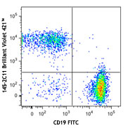
Brilliant Violet 421™ anti-mouse/human CD44

C57BL/6 mouse splenocytes were stained with CD44 (clone IM7)... -
Brilliant Violet 570™ anti-mouse/human CD44

C57BL/6 mouse splenocytes were stained with CD44 (clone IM7)... -
Brilliant Violet 785™ anti-mouse/human CD44

C57BL/6 mouse splenocytes were stained with CD44 (clone IM7)... -
Brilliant Violet 510™ anti-mouse/human CD44

C57BL/6 mouse splenocytes were stained with CD44 (clone IM7)... -
Ultra-LEAF™ Purified anti-mouse/human CD44

C57BL/6 mouse splenocytes stained with Ultra-LEAF™ purified ... -
Brilliant Violet 650™ anti-mouse/human CD44

C57BL/6 mouse splenocytes were stained with CD44 (clone IM7)... -
Purified anti-mouse/human CD44 (Maxpar® Ready)

Mouse splenocytes stained with 148Nd-anti-CD11b (M1/70) and ... -
Alexa Fluor® 594 anti-mouse/human CD44

bEND3 cells were fixed and blocked according to standard imm... 
Human paraffin-embedded intestine tissue was prepared with s... 
C57BL/6 mouse splenocytes were stained with CD44 (clone IM7)... -
PE/Dazzle™ 594 anti-mouse/human CD44

C57BL/6 mouse splenocytes were stained with CD44 (clone IM7)... -
Brilliant Violet 711™ anti-mouse/human CD44

C57BL/6 mouse splenocytes were stained with CD44 (clone IM7)... -
APC/Fire™ 750 anti-mouse/human CD44

C57BL/6 mouse splenocytes were stained with CD44 (clone IM7)... -
TotalSeq™-A0073 anti-mouse/human CD44
-
TotalSeq™-C0073 anti-mouse/human CD44
-
TotalSeq™-B0073 anti-mouse/human CD44
-
Spark YG™ 570 anti-mouse/human CD44

C57BL/6 mouse splenocytes were stained with CD44 (clone IM7)... 
Human paraffin-embedded intestine tissue slices were prepare... -
Spark YG™ 593 anti-mouse/human CD44

C57BL/6 splenocytes were stained with anti-mouse/human CD44 ... -
TotalSeq™-D0073 anti-mouse/human CD44
-
Brilliant Violet 750™ anti-mouse/human CD44

Mouse (C57BL/6) splenocytes stained with anti-mouse/human CD... -
PerCP/Fire™ 806 anti-mouse/human CD44

Human peripheral blood lymphocytes were stained with anti-hu... -
Spark Red™ 718 anti-mouse/human CD44

C57BL/6 mouse splenocytes were stained with anti -mouse/huma... -
Spark Blue™ 550 anti-mouse/human CD44 (Flexi-Fluor™)
-
Spark Red™ 718 anti-mouse/human CD44 (Flexi-Fluor™)


Follow Us