 Login/Register


- Clone
- MECA-79 (See other available formats)
- Regulatory Status
- RUO
- Other Names
- PNAd, Peripheral Node Addressin, MECA-79
- Isotype
- Rat IgM, κ
- Ave. Rating
- Submit a Review
- Product Citations
- publications

-

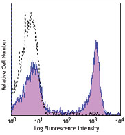
C57BL/6 mouse frozen lymph node section was fixed with 4% paraformaldehyde(PFA) for 10 minutes at room temperature and blocked with 5% FBS for 30 minutes at room temperature. Then the section was stained with 10 µg/ml of PNAd (clone MECA-79) purified, 5 µg/ml of CD45R/B220 (clone RA3-6B2) Alexa Fluor® 647 (green) overnight at 4°C, followed by 5 µg/ml of anti-rat IgM (clone MRM-47) Alexa Fluor® 594 (red) for 2 hours at room temperature. Nuclei were counterstained with DAPI (blue). The image was captured with a 10X objective. -

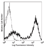
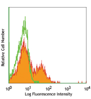
Human paraffin-embedded tonsil tissue slices were prepared with a standard protocol of deparaffination and rehydration. Antigen retrieval was done with Tris-Buffered Saline 1X (1.0M, pH7.4) at 95°C for 40 minutes. Tissue was washed with PBS/ 0.05% Tween20 twice for five minutes and blocked with 5% FBS and 0.2% gelatin for 30 minutes. Then, the tissue was stained with 10 µg/ml of purified anti-mouse/human PNAd antibody (clone MECA-79) at 4°C overnight. On the next day, the tissue was washed twice with PBS and stained with 2 µg/ml of biotin conjugated mouse anti-rat IgM secondary antibody for one hour at room temperature. After the incubation, the tissue was washed twice with PBS for five minutes and stained with Alexa Fluor® 488 Streptavidin (green) for 30 minutes at room temperature. Nuclei were counterstained with DAPI (blue). The image was captured with a 10X objective.
| Cat # | Size | Price | Quantity Check Availability | Save | ||
|---|---|---|---|---|---|---|
| 120801 | 50 µg | $71 | ||||
| 120802 | 500 µg | $262 | ||||
MECA-79 recognizes a carbohydrate epitope shared with a group of sulfation decorated sialomucins, including sulfated ligands for CD62L (CD34, GlyCAM-1, Sgp200, and a subset of MAdCAM-1). This set antigens has been referred to as peripheral node addressin (PNAd) with the molecular mass 50-250 kD. It has been identified that GlcNAc-6-SO4 sulfation contributes to MECA-79 binding and the core 1 β1,3-N-acetylglucosaminyltransferase is required for the generation of the MECA-79 epitope. MECA-79 is expressed on high endothelial venules (HEV) of lymphoid tissues, chronic inflamed tissues and rheumatoid synovia. The interaction of PNAd with CD62L receptor is involved in tethering and rolling of lymphocytes along HEV in lymphoid tissues. MECA-79 antibody reacts with mouse, human and many other species PNAd and blocks L-selectin-dependent lymphocyte adhesion in vitro and in vivo.
Product DetailsProduct Details
- Verified Reactivity
- Mouse, Human
- Antibody Type
- Monoclonal
- Host Species
- Rat
- Immunogen
- Mouse lymph node stromal cells
- Formulation
- Phosphate-buffered solution, pH 7.2, containing 0.09% sodium azide.
- Preparation
- The antibody was purified by affinity chromatography.
- Concentration
- 0.5 mg/ml
- Storage & Handling
- The antibody solution should be stored undiluted between 2°C and 8°C.
- Application
-
IHC-F - Quality tested
IHC-P - Verified
FC, IP, WB - Reported in the literature, not verified in house - Recommended Usage
-
Each lot of this antibody is quality control tested by immunohistochemical staining on frozen tissue sections. For immunohistochemistry, a concentration range of 2.5 - 10 µg/ml is suggested. For immunohistochemical staining on formalin-fixed paraffin-embedded tissue sections, the suggested use of this reagent is 5.0 - 10 µg/ml. It is recommended that the reagent be titrated for optimal performance for each application.
- Application Notes
-
Additional reported applications (for the relevant formats) include: in vitro and in vivo blocking1-4 of cell adhesion, immunohistochemical staining1,2,6 of acetone-fixed frozen sections and 4% paraformaldehyde-fixed paraffin-embedded sections, flow cytometry10,11, immunoprecipitation3,5, and Western blotting6,11.
-
Application References
(PubMed link indicates BioLegend citation) -
- Streeter PR, et al. 1988. J. Cell Biol. 107:1853. (IHC, Block)
- Michie SA, et al. 1993. Amer. J. Path. 143:1688. (IHC, Block)
- Berg EL, et al. 1991. J. Cell Biol. 114:343. (IP, Block)
- Berg EL, et al. 1993. Nature 366:695. (Block)
- Hemmerich S, et al. 1994. J. Exp. Med. 180:2219. (IP)
- Samulowitz U, et al. 2002. Am. J. Path. 160:1669. (IHC, WB)
- Burke SD, et al. 2007. Diabetes 56:2919. PubMed
- Hirakawa J, et al. 2010. J. Biol. Chem. 285:40864. PubMed
- Thomas SN, et al. 2012. J. Immunol. 189:2181. PubMed.
- Izawa D, et al. 1999. Int. Immunol. 11(12):1989-98. (FC, ICC)
- Sinha RK, et al. 2006. Immunology. 119(4):461-9. (FC, IHC-F, WB)
- Product Citations
-
- RRID
-
AB_493554 (BioLegend Cat. No. 120801)
AB_493555 (BioLegend Cat. No. 120802)
Antigen Details
- Structure
- Sulphated CD34, GlyCAM-1 and MAdCAM-1, Sgp200, sialomucin
- Distribution
-
High endothelial venules (HEV) in lymphoid tissues, inflamed tissues and rheumatoid synovia
- Function
- Leukocyte homing, rolling
- Ligand/Receptor
- CD62L
- Biology Area
- Cell Adhesion, Cell Biology, Immunology
- Molecular Family
- Adhesion Molecules
- Antigen References
-
1. Streeter PR, et al. 1988. J. Cell Biol. 107:1853.
2. Berg EL, et al. 1993. Nature 366:695.
3. Hemmerich S, et al. 1994. J. Exp. Med. 180:2219.
4. Mitoma J, et al. 2003. J. Biol. Chem. 278:9953.
5. Bruehl RE, et al. 2000. J. Biol. Chem. 275:32642.
6. Bistrup A, et al. 2004. Am. J. Pathol. 164:1635 - Gene ID
- 123803 View all products for this Gene ID 18203 View all products for this Gene ID
- UniProt
- View information about PNAd on UniProt.org

Related FAQs
Other Formats
View All PNAd Reagents Request Custom Conjugation| Description | Clone | Applications |
|---|---|---|
| Purified anti-mouse/human PNAd | MECA-79 | IHC-F,IHC-P,FC,IP,WB |
| Biotin anti-mouse/human PNAd | MECA-79 | IHC-F,IHC-P |
| Alexa Fluor® 594 anti-mouse/human PNAd | MECA-79 | IHC-P,IHC-F |
| Alexa Fluor® 647 anti-mouse/human PNAd | MECA-79 | IHC-P |
| Spark YG™ 570 anti-mouse/human PNAd Antibody | MECA-79 | IHC-F,IHC-P |
Customers Also Purchased
Compare Data Across All Formats
This data display is provided for general comparisons between formats.
Your actual data may vary due to variations in samples, target cells, instruments and their settings, staining conditions, and other factors.
If you need assistance with selecting the best format contact our expert technical support team.
-
Purified anti-mouse/human PNAd

C57BL/6 mouse frozen lymph node section was fixed with 4% pa... 
Human paraffin-embedded tonsil tissue slices were prepared w... -
Biotin anti-mouse/human PNAd

C57BL/6 mouse frozen lymph node section was fixed with 4% pa... 
Human paraffin-embedded tonsil tissue slices were prepared w... -
Alexa Fluor® 594 anti-mouse/human PNAd

Human paraffin-embedded tonsil tissue slices were prepared w... 
C57BL/6 mouse frozen lymph node section was fixed with 4% pa... -
Alexa Fluor® 647 anti-mouse/human PNAd

Human paraffin-embedded tonsil tissue slices were prepared w... -
Spark YG™ 570 anti-mouse/human PNAd Antibody

Human paraffin-embedded tonsil tissue slices were prepared w... 
C57BL/6 mouse frozen lymph node section was fixed with 4% pa...


Follow Us