 Login/Register


- Clone
- A21001C (See other available formats)
- Regulatory Status
- RUO
- Other Names
- Protein kinase B alpha, PRK-BA, PKB, Serine/Threonine specific kinase RAC alpha, Protein kinase Akt, RAC
- Isotype
- Mouse IgG1, κ
- Ave. Rating
- Submit a Review
- Product Citations
- publications

-

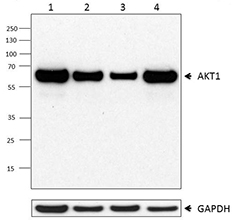
Whole cell extracts (15 µg total protein) from serum-starved HeLa cells untreated (-) or treated (+) with IGF-1 (100 ng/ml, 30 min), and from serum-starved NIH/3T3 cells untreated (-) or treated (+) with PDGF-BB (200 ng/mL, 15 min) were resolved on a 4-12% Bis-Tris gel, transferred to a PVDF membrane, and probed with 1 µg/mL (1:500 dilution) of purified anti-AKT Phospho (Ser473) (clone A21001C) overnight at 4°C. Proteins were visualized by chemiluminescence detection using HRP goat anti-mouse IgG (Cat. No. 405306) at a 1:3000 dilution. Equal AKT Phospho (Ser473) loading was confirmed by probing membranes with anti-pan AKT1 (clone O94E10) (Cat. No. 680302) at a 1:500 dilution. Direct-Blot™ HRP anti-GAPDH (Cat. No. 607904) was used as a loading control at a 1:50000 dilution. Western-Ready™ ECL Substrate Plus Kit (Cat. No. 426317) was used as a detection agent. Lane M: Molecular weight marker -

Untreated Jurkat cells (positive control, panel A) and Jurkat cells treated with 1 µM wortmannin for 2 hours (negative control, panel B) were fixed with 4% paraformaldehyde for 10 minutes, permeabilized with ice cold MeOH for 10 minutes, and blocked with 5% FBS for 60 minutes. Cells were then intracellularly stained with 5.0 µg/mL purified anti-AKT Phospho (Ser473) (clone A21001C) overnight at 4°C, followed by incubation with Alexa Fluor® 594 goat anti-mouse IgG (Cat. No. 405326) at 2.0 µg/mL. Nuclei were counterstained with DAPI and the image was captured with a 60X objective. Scale: 50 µm -

Jurkat cells untreated (positive target, filled histogram), and treated with 1.0 µM wortmannin for 2 hours (negative target, solid line, open histogram) were fixed and permeabilized using the True-Phos™ Fix/Perm Buffer Set (Cat. No. 425401) and intracellularly stained with either purified anti-AKT Phospho (Ser473) (clone A21001C) (solid line histograms) or purified mouse IgG1, κ isotype control (dashed line, open histogram) followed by PE goat anti-mouse IgG.
| Cat # | Size | Price | Quantity Check Availability | Save | ||
|---|---|---|---|---|---|---|
| 606551 | 25 µg | $118 | ||||
| 606552 | 100 µg | $347 | ||||
AKT (also known as protein kinase B alpha) is a 60 kD serine/threonine-specific kinase containing a pleckstrin domain. AKT plays a critical role in controlling survival and apoptosis. This kinase is ubiquitously expressed and translocates to the membrane upon cell activation. It is activated by insulin and various growth and survival factors to function in a wortmannin-sensitive pathway involving PI 3-kinase. AKT is activated by phospholipid binding and activation loop phosphorylation at Thr308 by PDK1 and by phosphorylation within the carboxy terminus at Ser473. The previously elusive PDK2, responsible for phosphorylation of AKT at Ser473, has been identified as a mammalian target of rapamycin (mTOR) in a rapamycin-insensitive complex with rictor and Sin1. Activated AKT phosphorylates a wide range of substrates including transcription factors (e.g. FOXO1), kinases (GSK-3beta, Raf-1, ASK, Chk1) and other proteins with important signaling roles (e.g. Bad, MDM2). AKT promotes cell survival by inhibiting apoptosis through phosphorylation of several targets, including Bad, forkhead transcription factor, c-Raf and caspase-9. AKT also plays a critical role in cell growth by directly phosphorylating mTOR in a rapamycin-sensitive complex containing raptor. More importantly, AKT phosphorylates and inactivates tuberin (TSC2), an inhibitor of mTOR within the mTOR-raptor complex.
Product DetailsProduct Details
- Verified Reactivity
- Human, Mouse
- Antibody Type
- Monoclonal
- Host Species
- Mouse
- Immunogen
- Synthetic peptide of human AKT phosphorylated at Ser473
- Formulation
- Phosphate-buffered solution, pH 7.2, containing 0.09% sodium azide
- Preparation
- The antibody was purified by affinity chromatography.
- Concentration
- 0.5 mg/mL
- Storage & Handling
- The antibody solution should be stored undiluted between 2°C and 8°C.
- Application
-
WB - Quality tested
ICC, ICFC - Verified - Recommended Usage
-
Each lot of this antibody is quality control tested by western blotting. For western blotting, the suggested use of this reagent is 0.25 - 1.0 µg/mL. For immunocytochemistry, a concentration range of 2.5 - 5.0 μg/mL is recommended. For flow cytometric staining, the suggested use of this reagent is ≤ 0.03 µg per million cells in 100 µL volume. It is recommended that the reagent be titrated for optimal performance for each application.
- Application Notes
-
This clone was tested by Western Blot using lysates prepared from HeLa cells untreated or treated with IGF-1, and NIH/3T3 cell untreated or treated with PDGF.
This clone is suitable for ICC in cells treated with PFA and permeabilized with either methanol or Triton X-100. - RRID
-
AB_2924618 (BioLegend Cat. No. 606551)
AB_2924618 (BioLegend Cat. No. 606552)
Antigen Details
- Structure
- AKT is a 480 amino acid protein with a predicted molecular weight of 56 kD
- Distribution
-
Ubiquitously expressed, translocates to the membrane upon activation
- Function
- Catalytically inactive multimeric complex. AKT is activated by phospholipid binding and activation loop phosphorylation at Thr308 by PDK1 and by phosphorylation within the carboxy terminus at Ser473 by PDK2.
- Interaction
- Interacts through pleckstrin homology domain with second messengers. Interacts with AKTIP, CDKN1B.
- Biology Area
- Cell Biology, Cell Cycle/DNA Replication, Cell Death
- Molecular Family
- Phospho-Proteins, Protein Kinases/Phosphatase
- Antigen References
-
- Jacinto E, et al. 2006. Cell. 127:125-37.
- Inoki K, et al. 2002. Nat Cell Biol. 4:648-57.
- Franke TF, et al. 1995. Cell. 81:727-36.
- Brunet A, et al. 1999. Cell. 96:857-68.
- Yang WL, et al. 2010. Cell Cycle. 9:487-97.
- Gene ID
- 207 View all products for this Gene ID
- UniProt
- View information about AKT Phospho Ser473 on UniProt.org

Related FAQs
Other Formats
View All AKT Phospho Ser473 Reagents Request Custom Conjugation| Description | Clone | Applications |
|---|---|---|
| Purified anti-AKT Phospho (Ser473) | A21001C | WB,ICC,ICFC |
| Alexa Fluor® 647 anti-AKT Phospho (Ser473) | A21001C | ICFC |
| PE anti-AKT Phospho (Ser473) | A21001C | ICFC |
Customers Also Purchased
Compare Data Across All Formats
This data display is provided for general comparisons between formats.
Your actual data may vary due to variations in samples, target cells, instruments and their settings, staining conditions, and other factors.
If you need assistance with selecting the best format contact our expert technical support team.
-
Purified anti-AKT Phospho (Ser473)

Whole cell extracts (15 µg total protein) from serum-starved... 
Untreated Jurkat cells (positive control, panel A) and Jurka... 
Jurkat cells untreated (positive target, filled histogram), ... -
Alexa Fluor® 647 anti-AKT Phospho (Ser473)

Jurkat cells untreated (positive control, filled histogram),... -
PE anti-AKT Phospho (Ser473)

Jurkat cells untreated (positive control, filled histogram),...


Follow Us