 Login/Register


- Clone
- H11 (See other available formats)
- Regulatory Status
- RUO
- Other Names
- MHC
- Previously
-
Covance Catalog# MMS-460R
- Isotype
- Mouse IgM, κ
- Ave. Rating
- Submit a Review
- Product Citations
- publications

-

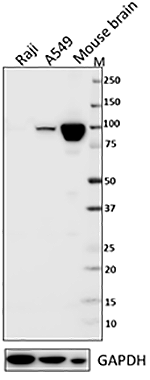
Total cell lysates (15 µg total protein) from A431, HaCat, HeLa, U937, and NTERA-2 cells were resolved by 4-12% Bis-Tris gel electrophoresis, transferred to a nitrocellulose membrane, and probed with a 1:2500 dilution of Purified anti-Myosin Heavy Chain Antibody, clone H11, overnight at 4°C. Proteins were visualized by chemiluminescence detection using HRP goat anti-mouse IgG Antibody (Cat. No. 405306) at a 1:3000 dilution. Direct-Blot™ HRP anti-GAPDH Antibody (Cat. No. 607904) was used as a loading control at a 1:50000 dilution (lower). Lane M: Molecular Weight marker. -

Immunofluorescence of HUVEC cells with (A) mouse isotype control (negative) or (B-D) myosin heavy chain antibody (clone H11). Alexa Fluor® 488 (green) Goat anti-mouse IgG (Cat. No. 405319) was used as secondary antibody. Nuclei were counterstained with DAPI (Blue, Cat. No. 422801). The image was captured with a 60X objective using KEYENCE BZ-X700 fluorescence microscope. Exposure time (seconds) for (A) is 1/10, and (B-D) is 1/15. Concentrations for (A, B) is 1/100, (C) is 1/250 and (D) is 1/500.
| Cat # | Size | Price | Quantity Check Availability | Save | ||
|---|---|---|---|---|---|---|
| 922701 | 100 µL | $256 | ||||
Myosin heavy chain is an actin-based motor protein with an approximate molecular weight of 223 kD. Myosin heavy chain can be split into 1 light meromysin and 1 heavy meromysin chain (and further into 2 globular subfragments and 1 rod-shaped subfragment). This protein is highly expressed in muscle. Motor proteins that convert chemical energy derived from ATP hydrolysis into mechanical force that drives motile processes such as cytokinesis, vesicular transport, and cellular locomotion. Myosin heavy chain is modified by acetylation in cysteine residues in the S1 domain for myosin ATPase activity. Muscle myosin is a hexameric protein consisting of 2 heavy chain subunits and 2 alkali light chain subunits and 2 regulatory light chain subunits that forms filaments.
Product DetailsProduct Details
- Verified Reactivity
- Human
- Antibody Type
- Monoclonal
- Host Species
- Mouse
- Immunogen
- This antibody was generated against a complex mixture of phosphoproteins extracted from Madin-Darby Canine Kidney Cells.
- Preparation
- Ascites
- Concentration
- The concentration is not quantified as this product is sold as undiluted crude mouse ascites fluid. The concentration might vary from lot-to-lot and an estimated concentration would be 1-3 mg/ml.
- Storage & Handling
- Store at -20°C. Upon initial thawing, apportion into working aliquots and store at -20°C. Avoid repeated freeze-thaw cycles to prevent denaturing the antibody. For long-term storage, keep the antibody at -80°C.
- Application
-
WB - Quality tested
ICC - Verified - Recommended Usage
-
Each lot of this antibody is quality control tested by Western blotting. For Western blotting, the suggested use of this reagent is a dilution of 1:1000 - 1:2500. For immunocytochemistry, a dilution of 1:1000 - 1:250 is recommended. For immunoprecipitation, the suggested use of this reagent is a dilution of 1:150. It is recommended that the reagent be titrated for optimal performance for each application.
- Application Notes
-
This antibody is effective in immunoblotting (WB), immunofluorescence (ICC) and immunoprecipitation (IP).
*Predicted MW = 205 kD
**Recommend using extracts prepared in buffers containing 0.5% Triton X-100 and 0.5% sodium deoxycholate.
This antibody binds specifically to denatured as well as native myosin heavy chain extracts and will not react with amphibian myosins. -
Application References
(PubMed link indicates BioLegend citation) -
- Chew TL, et al. 2002. J. Cell Biol. 156:543. (ICC) PubMed
- Warren SL, et al. 1988. Mol Cell Biol. 8:632.
- Product Citations
-
- RRID
-
AB_2565417 (BioLegend Cat. No. 922701)
Antigen Details
- Structure
- Actin-based motor protein, approximately 223 kD. Myosin heavy chain can be split into 1 light meromysin and 1 heavy meromysin chain (and further into 2 globular subfragments and 1 rod-shaped subfragment).
- Distribution
-
Highly expressed in muscle.
- Function
- Motor proteins that convert chemical energy derived from ATP hydrolysis into mechanical force that drives motile processes such as cytokinesis, vesicular transport, and cellular locomotion.
- Interaction
- Muscle myosin is a hexameric protein consisting of 2 heavy chain subunits and 2 alkali light chain subunits and 2 regulatory light chain subunits. Forms filaments.
- Modification
- Alkylation.
- Biology Area
- Cell Biology, Cell Motility/Cytoskeleton/Structure, Neuroscience, Neuroscience Cell Markers
- Antigen References
-
1. Weiss A, et al. 1999. P. Natl. Acad. Sci. USA 96:2598.
2. Schiaffino S, et al. 1996. Physiol. Rev. 76:371 (review) - Regulation
- Cysteine residues in the S1 domain are alkylated for myosin ATPase activity.
- Gene ID
- NA
- UniProt
- View information about Myosin Heavy Chain on UniProt.org
Related Pages & Pathways
Pathways


Related FAQs
Other Formats
View All Myosin Heavy Chain Reagents Request Custom Conjugation| Description | Clone | Applications |
|---|---|---|
| Anti-Myosin Heavy Chain | H11 | WB,ICC |
| Purified anti-Myosin Heavy Chain | H11 | WB,ICC |
Customers Also Purchased
Compare Data Across All Formats
This data display is provided for general comparisons between formats.
Your actual data may vary due to variations in samples, target cells, instruments and their settings, staining conditions, and other factors.
If you need assistance with selecting the best format contact our expert technical support team.
-
Anti-Myosin Heavy Chain

Immunofluorescence of HUVEC cells with (A) mouse isotype con... 
Total cell lysates (15 µg total protein) from A431, HaCat, H... -
Purified anti-Myosin Heavy Chain

Total cell lysates (15 µg total protein) from the indicated ... 
ICC staining of purified mouse IgM, κ isotype ctrl antibody ...


Follow Us