 Login/Register


- Clone
- DECMA-1 (See other available formats)
- Regulatory Status
- RUO
- Other Names
- E-Cadherin, Cadherin-1, CDH1, and UVO
- Isotype
- Rat IgG1, κ
- Ave. Rating
- Submit a Review
- Product Citations
- publications

-

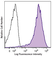
MDCK epithelial cell line was stained with CD324 (clone DECMA-1) Alexa Fluor® 647 (filled histogram) or rat IgG1, κ Alexa Fluor® 647 isotype control (open histogram). -

Madin-darby canine kidney epithelial cell line, MDCK, was cultured in a chamber slide until confluent. The cells were fixed with 1% paraformaldehyde (PFA) for 10 minutes, permeabilized with 0.5% Triton X-100 for 10 minutes, and blocked with 5% FBS for 30 minutes. The cells were then intracellularly stained with 5 µg/mL of CD324/E-Cadherin (clone DECMA-1) Alexa Fluor® 647 (red) in blocking buffer overnight at 4°C. Nuclei were counterstained with DAPI (blue). The image was captured with 40X objective. -

C57BL/6 mouse frozen intestine section was fixed with 4% paraformaldehyde (PFA) for 10 minutes, permeabilized with 0.5% Triton X-100 for ten minutes, and blocked with 5% FBS for 30 minutes at room temperature. Then the section was stained with 5 µg/mL CD324 (clone DECMA-1) Alexa Fluor® 647 (red) and Ki-67 (clone 11F6) Alexa Fluor® 594 (green) overnight at 4°C. Nuclei were counterstained with DAPI (blue). The image was captured by 10X objective. -

Paraformaldehyde-fixed (4%), 500 μm-thick mouse lung tissue section was processed according to the Ce3DTM Tissue Clearing Kit protocol (cat. no. 427701). The section was costained with anti-mouse CD45 Antibody (clone 30-F11) Alexa Fluor® 488 at 5 μg/mL (green), anti-mouse/human CD45R/B220 Antibody (clone RA3-6B2) Alexa Fluor® 594 at 5 μg/mL (yellow), and anti-mouse/human CD324 (E-Cadherin) Antibody (clone DECMA-1) Alexa Fluor® 647 at 5 μg/mL (magenta) and counterstained with DAPI (blue). The section was then optically cleared and mounted in a sample chamber. The image was captured with a 20X objective using Zeiss 780 confocal microscope and processed by Imaris image analysis software.
Watch the video. -

Confocal image of C57BL/6 mouse liver sample acquired using the IBEX method of highly multiplexed antibody-based imaging: E-Cadherin (red) in Cycle 1 and CD45 (cyan) in Cycle 3. Tissues were prepared using ~1% (vol/vol) formaldehyde and a detergent. Following fixation, samples are immersed in 30% (wt/vol) sucrose for cryoprotection. Images are courtesy of Drs. Andrea J. Radtke and Ronald N. Germain of the Center for Advanced Tissue Imaging (CAT-I) in the National Institute of Allergy and Infectious Diseases (NIAID, NIH). -

Confocal image of C57BL/6 mouse liver sample acquired using the IBEX method of highly multiplexed antibody-based imaging: E-Cadherin (blue) in Cycle 1 and MHCII (magenta) in Cycle 4. Tissues were prepared using ~1% (vol/vol) formaldehyde and a detergent. Following fixation, samples are immersed in 30% (wt/vol) sucrose for cryoprotection. Images are courtesy of Drs. Andrea J. Radtke and Ronald N. Germain of the Center for Advanced Tissue Imaging (CAT-I) in the National Institute of Allergy and Infectious Diseases (NIAID, NIH).
| Cat # | Size | Price | Quantity Check Availability | Save | ||
|---|---|---|---|---|---|---|
| 147307 | 25 µg | $124 | ||||
| 147308 | 100 µg | $323 | ||||
CD324, also known as E-cadherin, cadherin-1, CDH1, and UVO is a member of the cadherin superfamily. It is a calcium-dependent, transmembrane cell-cell adhesion glycoprotein composed of four extracellular cadherin repeats and a highly conserved cytoplasmic tail region. CD324 is widely expressed in epithelial cells in the colon, uterus, liver, keratinocytes, brain, heart, muscle, kidney, and pancreas as well as erythroid cells. CD324 functions as a cell adhesion molecule involved in development, bacterial pathogenesis, and tumor invasion. In bacterial pathogenesis, the ectodomain of CD324 mediates bacterial adhesion to mammalian cells, while the cytoplasmic domain is required for internalization. CD324 binds to the αEβ7 integrin to mediate cell adhesion and also interacts with a number of intracellular proteins including including erbin, ezrin, caspase-3, caspase-8, β-catenin, presenilin 1, and casein kinase II as well as other extracellular proteins including the EGF receptor.
Product DetailsProduct Details
- Verified Reactivity
- Mouse, Human
- Reported Reactivity
- Cynomolgus, Dog, Pig
- Antibody Type
- Monoclonal
- Host Species
- Rat
- Immunogen
- E-Cadherin extracellular domain
- Formulation
- Phosphate-buffered solution, pH 7.2, containing 0.09% sodium azide.
- Preparation
- The antibody was purified by affinity chromatography and conjugated with Alexa Fluor® 647 under optimal conditions.
- Concentration
- 0.5 mg/mL
- Storage & Handling
- The antibody solution should be stored undiluted between 2°C and 8°C, and protected from prolonged exposure to light. Do not freeze.
- Application
-
FC - Quality tested
ICC, IHC-F, 3D IHC - VerifiedSB - Reported in the literature, not verified in house
- Recommended Usage
-
Each lot of this antibody is quality control tested by immunofluorescent staining with flow cytometric analysis. For flow cytometric staining, the suggested use of this reagent is ≤ 1.0 µg per million cells in 100 µL volume. For immunocytochemistry, a concentration range of 2.0 - 10 μg/mL is recommended. For immunohistochemical staining on frozen tissue sections, a concentration range of 5.0 - 10 µg/mL is suggested. For 3D immunohistochemistry on formalin-fixed tissues, a concentration of 5.0 µg/mL is suggested. It is recommended that the reagent be titrated for optimal performance for each application.
It is also recommended using EDTA-based solutions for dissociating attachment-dependent cell lines.
* Alexa Fluor® 647 has a maximum emission of 668 nm when it is excited at 633 nm / 635 nm.
Alexa Fluor® and Pacific Blue™ are trademarks of Life Technologies Corporation.
View full statement regarding label licenses - Excitation Laser
-
Red Laser (633 nm)
- Application Notes
-
Additional reported applications (for relevant formats) include: immunoprecipitation1, Western Blotting1, immunomicroscopy3, biological function1,2, and spatial biology (IBEX)4,5.
- Additional Product Notes
-
Iterative Bleaching Extended multi-pleXity (IBEX) is a fluorescent imaging technique capable of highly-multiplexed spatial analysis. The method relies on cyclical bleaching of panels of fluorescent antibodies in order to image and analyze many markers over multiple cycles of staining, imaging, and, bleaching. It is a community-developed open-access method developed by the Center for Advanced Tissue Imaging (CAT-I) in the National Institute of Allergy and Infectious Diseases (NIAID, NIH).
-
Application References
(PubMed link indicates BioLegend citation) -
- Vestweber D, et al. 1985. EMBO. 4:3393. (IP, WB, FA)
- Nakagawa M, et al. 2001. J. Cell Sci. 114:1829. (FA in canine cells)
- Mohamet L, et al. 2010. PLoS ONE. 5:e12921. (IF)
- Radtke AJ, et al. 2020. Proc Natl Acad Sci U S A. 117:33455-65. (SB) PubMed
- Radtke AJ, et al. 2022. Nat Protoc. 17:378-401. (SB) PubMed
- Product Citations
-
- RRID
-
AB_2563954 (BioLegend Cat. No. 147307)
AB_2563955 (BioLegend Cat. No. 147308)
Antigen Details
- Structure
- Member of the cadherin superfamily. Calcium-dependent, transmembrane cell-cell adhesion glycoprotein composed of four extracellular cadherin repeats and a highly conserved cytoplasmic tail region.
- Distribution
-
Widely expressed in epithelial cells in the colon, uterus, liver, keratinocytes, brain, heart, muscle, kidney, and pancreas as well as erythroid cells.
- Function
- Cell adhesion molecule involved in development, bacterial pathogenesis, and tumor invasion. The ectodomain of CD324 mediates bacterial adhesion to mammalian cells, while the cytoplasmic domain is required for internalization.
- Interaction
- Interacts with a variety of proteins including erbin, ezrin, caspase-3, caspase-8, EGF receptor, β-catenin, presenilin 1, casein kinase II, and others.
- Ligand/Receptor
- αEβ7 integrin.
- Cell Type
- Embryonic Stem Cells
- Biology Area
- Cell Adhesion, Cell Biology, Immunology, Innate Immunity, Neuroscience, Stem Cells, Synaptic Biology
- Molecular Family
- Adhesion Molecules, CD Molecules
- Antigen References
-
1. Overduin M, et al. 1995. Science 267:386.
2. Boggon TJ, et al. 2002. Science 296:1308.
3. Berx G, et al. 1995. EMBO J. 14:6107.
4. Perl AK, et al. 1998. Nature 392:190. - Gene ID
- 999 View all products for this Gene ID 12550 View all products for this Gene ID
- UniProt
- View information about CD324 on UniProt.org
Related Pages & Pathways
Pathways


Related FAQs
- If an antibody clone has been previously successfully used in IBEX in one fluorescent format, will other antibody formats work as well?
-
It’s likely that other fluorophore conjugates to the same antibody clone will also be compatible with IBEX using the same sample fixation procedure. Ultimately a directly conjugated antibody’s utility in fluorescent imaging and IBEX may be specific to the sample and microscope being used in the experiment. Some antibody clone conjugates may perform better than others due to performance differences in non-specific binding, fluorophore brightness, and other biochemical properties unique to that conjugate.
- Will antibodies my lab is already using for fluorescent or chromogenic IHC work in IBEX?
-
Fundamentally, IBEX as a technique that works much in the same way as single antibody panels or single marker IF/IHC. If you’re already successfully using an antibody clone on a sample of interest, it is likely that clone will have utility in IBEX. It is expected some optimization and testing of different antibody fluorophore conjugates will be required to find a suitable format; however, legacy microscopy techniques like chromogenic IHC on fixed or frozen tissue is an excellent place to start looking for useful antibodies.
- Are other fluorophores compatible with IBEX?
-
Over 18 fluorescent formats have been screened for use in IBEX, however, it is likely that other fluorophores are able to be rapidly bleached in IBEX. If a fluorophore format is already suitable for your imaging platform it can be tested for compatibility in IBEX.
- The same antibody works in one tissue type but not another. What is happening?
-
Differences in tissue properties may impact both the ability of an antibody to bind its target specifically and impact the ability of a specific fluorophore conjugate to overcome the background fluorescent signal in a given tissue. Secondary stains, as well as testing multiple fluorescent conjugates of the same clone, may help to troubleshoot challenging targets or tissues. Using a reference control tissue may also give confidence in the specificity of your staining.
- How can I be sure the staining I’m seeing in my tissue is real?
-
In general, best practices for validating an antibody in traditional chromogenic or fluorescent IHC are applicable to IBEX. Please reference the Nature Methods review on antibody based multiplexed imaging for resources on validating antibodies for IBEX.
Other Formats
View All CD324 Reagents Request Custom Conjugation| Description | Clone | Applications |
|---|---|---|
| Purified anti-mouse/human CD324 (E-Cadherin) | DECMA-1 | FC,ICC,IP,WB,FA |
| PE anti-mouse/human CD324 (E-Cadherin) | DECMA-1 | FC |
| Alexa Fluor® 594 anti-mouse/human CD324 (E-Cadherin) | DECMA-1 | ICC,IHC-P,IHC-F,3D IHC |
| Alexa Fluor® 647 anti-mouse/human CD324 (E-Cadherin) | DECMA-1 | FC,ICC,IHC-F,3D IHC,SB |
| PE/Cyanine7 anti-mouse/human CD324 (E-Cadherin) | DECMA-1 | FC |
| PE/Dazzle™ 594 anti-mouse/human CD324 (E-Cadherin) | DECMA-1 | FC |
| PerCP/Cyanine5.5 anti-mouse/human CD324 (E-Cadherin) | DECMA-1 | FC |
| APC anti-mouse/human CD324 (E-Cadherin) | DECMA-1 | FC |
| Brilliant Violet 421™ anti-mouse/human CD324 (E-Cadherin) | DECMA-1 | FC |
| APC/Fire™ 750 anti-mouse/human CD324 (E-Cadherin) | DECMA-1 | FC |
Customers Also Purchased
Compare Data Across All Formats
This data display is provided for general comparisons between formats.
Your actual data may vary due to variations in samples, target cells, instruments and their settings, staining conditions, and other factors.
If you need assistance with selecting the best format contact our expert technical support team.
-
Purified anti-mouse/human CD324 (E-Cadherin)

Canine kidney cell line MDCK was cultured in a chamber slide... 
MDCK epithelial cell line was stained with purified CD324 (c... -
PE anti-mouse/human CD324 (E-Cadherin)

MDCK epithelial cell line was stained with CD324 (clone DECM... -
Alexa Fluor® 594 anti-mouse/human CD324 (E-Cadherin)

Madin-darby canine kidney epithelial cell line, MDCK, was cu... 
C57BL/6 mouse frozen intestine section was fixed with 4% par... 
Human paraffin-embedded colon cancer tissue slices were prep... 
Paraformaldehyde-fixed (1%), 500 μm-thick mouse liver tissue... -
Alexa Fluor® 647 anti-mouse/human CD324 (E-Cadherin)

MDCK epithelial cell line was stained with CD324 (clone DECM... 
Madin-darby canine kidney epithelial cell line, MDCK, was cu... 
C57BL/6 mouse frozen intestine section was fixed with 4% par... 
Paraformaldehyde-fixed (4%), 500 μm-thick mouse lung tissue ... 
Confocal image of C57BL/6 mouse liver sample acquired using ... 
Confocal image of C57BL/6 mouse liver sample acquired using ... -
PE/Cyanine7 anti-mouse/human CD324 (E-Cadherin)

MDCK epithelial cell line was stained with CD324 (clone DECM... -
PE/Dazzle™ 594 anti-mouse/human CD324 (E-Cadherin)

MDCK epithelial cell line was stained with CD324 (E-Cadherin... -
PerCP/Cyanine5.5 anti-mouse/human CD324 (E-Cadherin)

MDCK epithelial cell line was stained with CD324 (E-Cadherin... -
APC anti-mouse/human CD324 (E-Cadherin)

MDCK epithelial cell line was stained with CD324 (clone DECM... -
Brilliant Violet 421™ anti-mouse/human CD324 (E-Cadherin)

MDCK epithelial cell line was stained with CD324 (E-Cadherin... -
APC/Fire™ 750 anti-mouse/human CD324 (E-Cadherin)

MDCK epithelial cell line was stained with CD324 (clone DECM...


Follow Us