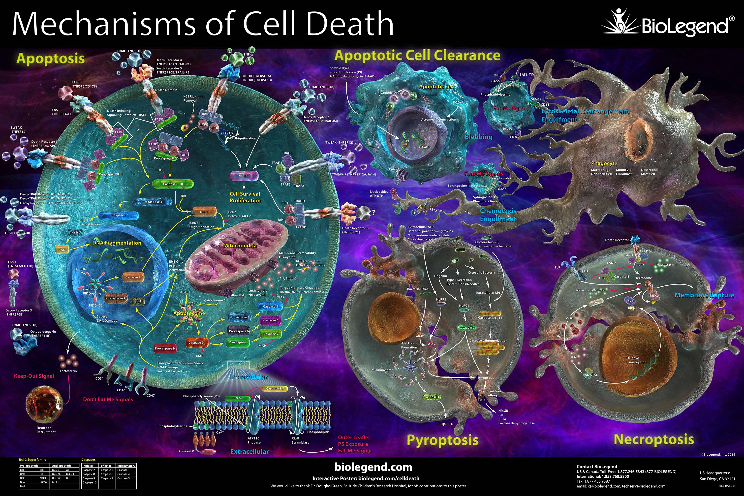
Live/Dead Indicators
Within a sample, it can be important to assess the viability and general health of a cell. Dying/dead cells can create debris that lead to false positive staining within a flow cytometry experiment. A majority of the reagents in this category function on the concept that intact membranes of living cells help to limit the binding of a fluorescent dye or chemical probe. Learn about each category of our non-antibody chemical probes below to help decide which reagent is ideal for your assay.
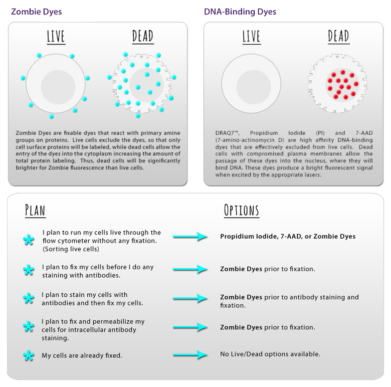
Fixable Live/Dead Indicators
Fixation and permeabilization reagents (such as alcohols and paraformaldehyde) can unwind and denature DNA, dislodging DNA-binding dyes and giving false negative results. Simultaneously, free dye could find its way into previously healthy cells that have now been fixed/permeabilized and give a false positive result.
To avoid this issue, we provide Zombie Dyes, which bind the amine groups present on proteins. While live cells are stained to a low degree due to surface proteins, dead cells will stain much more brightly due to the high abundance of internal proteins that can be stained once membranes are compromised.
Learn about Zombie Dyes >
Non-Fixable Live/Dead Indicators
One of the earliest techniques for identifying dead cells involved the use of nucleic acid-binding dyes such as Propidium Iodide, 7-AAD, DAPI, and BioLegend’s Helix NP™ (non-permeant) dyes. These dyes work on the principle that live and healthy cells possess intact membranes that prevent these dyes from gaining access to the nucleus. However, dying cells will have compromised membranes that allow these dyes to bind directly to the DNA.
It should be noted that fixation and permeabilization procedures can cause potential issues with the ability of these dyes to remain bound to their target.
View Our DNA-Binding Dyes >
 Login / Register


Follow Us