 Login/Register


Brilliant Violet™ antibody conjugates, proudly co-developed by BioLegend and Sirigen, are an innovative class of novel research reagents, providing more options for your multicolor flow cytometry panels and getting you better results. Maximize the capacity of your violet laser with our large selection of directly labeled Brilliant Violet™ antibody conjugates.
Loading...
Brilliant Violet™
Features
|
• Extremely Bright |
• Nontoxic - for Sorting or Live Cell Imaging |
Brilliant Violet™ Family
Brilliant Violet 421™ | Brilliant Violet 510™ | Brilliant Violet 570™ | Brilliant Violet 605™ | Brilliant Violet 650™ | Brilliant Violet 711™ | Brilliant Violet 750™ | Brilliant Violet 785™
Brilliant Violet 421™
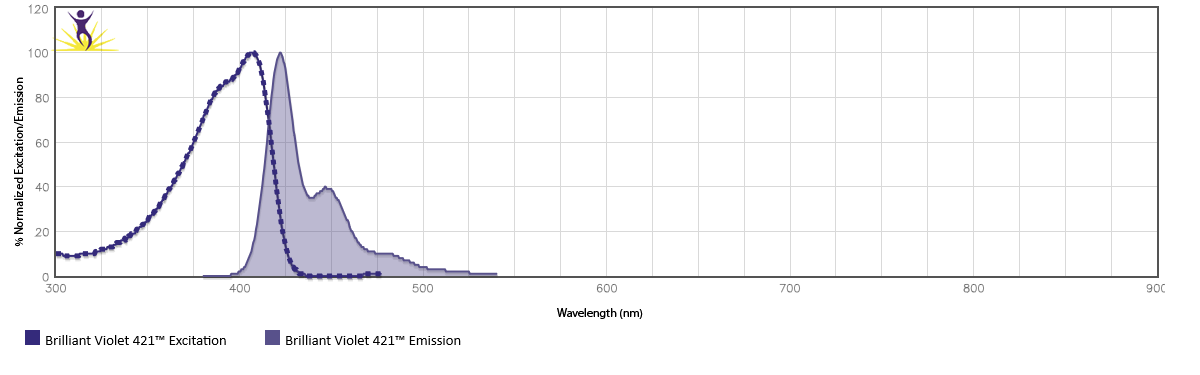 The first in the series, BV421™ can increase assay sensitivity by logarithmic orders of magnitude without increasing background or spillover, making it ideal for detecting rare cell populations or weakly expressed cell markers. It is exceptionally photostable, enabling the visualization of antigens with directly conjugated antibodies for confocal microscopy applications. Est. MW = 70 kD.
Excitation Max = 405 nm, Emission Max = 421 nm
Recommended filter = 450/50
Comparable Fluorophores: Pacific Blue™, Alexa Fluor® 405, eFluor® 450, BD Horizon™ V450, Cascade Blue™
Brightness = 5 (On a scale from 1 to 5, with 5 being the brightest.)
Molar Extinction Coeff. = 2,500,000 M-1cm-1
Quantum Yield = 0.65 in DPBS
Beta Testing -
- Resolution of mouse NK1.1 populations
- High sensitivity for mouse CD127
- Non-toxic for cell sorting - mouse CD4
- Multi-color microscopy with human CD56
- Resolution of human CD56 populations
- Resolution of human CD45RA v RO
Brilliant Violet 510™
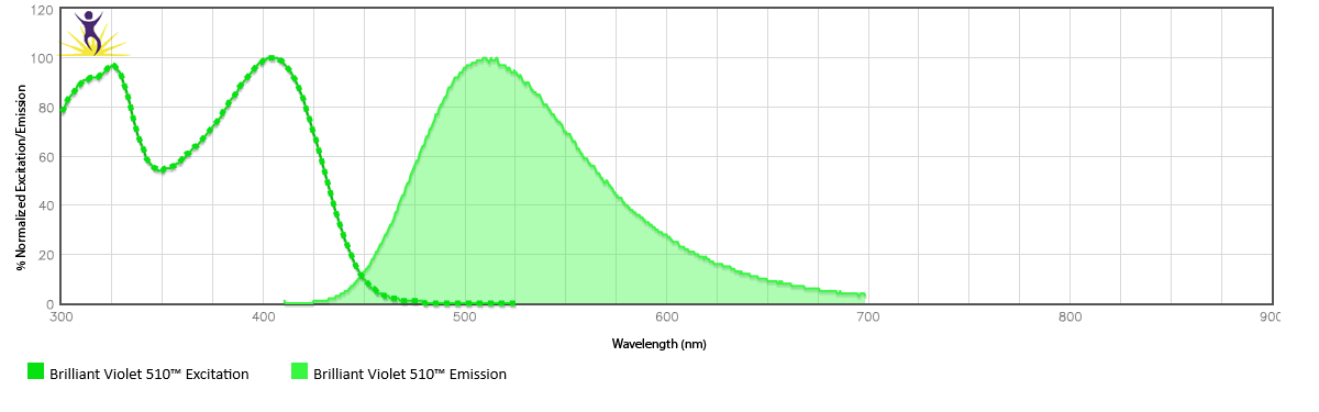
Brilliant Violet 510™ is a novel non-tandem polymer with excellent signal-to-noise, excited by the violet laser. It can provide dramatic improvements over existing fluorophores emitting in this range such as Pacific Orange™, AmCyan, and Horizon™ V500. Est. MW = 77 kD.
Excitation Max = 405 nm, Emission Max = 510 nm
Recommended filter = 510/50
Comparable Fluorophores: BD Horizon™ V500, Pacific Orange™, Cascade Yellow™, AmCyan
Brightness = 2 (On a scale from 1 to 5, with 5 being the brightest.)
Molar Extinction Coeff. = 577,000 M-1cm-1
Quantum Yield = 0.44 in DPBS
Beta Testing -
- Cytokine profiling of T cell subsets using Brilliant Violet™ fluorophores
- 15 Color Assay using Brilliant Violet™ Fluorophores
Brilliant Violet 570™
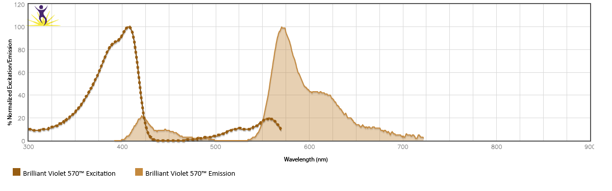
Brilliant Violet 570™ is a novel molecule based on the Brilliant Violet 421™ polymer core. It provides a much brighter alternative to Pacific Orange™ for multicolor flow on the violet laser and is a better alternative to nanocrystals for intracellular flow cytometry. Est. MW = 60 kD.
Excitation Max = 405 nm, Emission Max = 570 nm
Recommended filter = 585/42
Comparable Fluorophores: Pacific Orange™, Cascade Yellow™, Qdot® 545, Qdot® 565, eFluor® 565NC
Brightness = 2 (On a scale from 1 to 5, with 5 being the brightest.)
Molar Extinction Coeff. = 2,300,000 M-1cm-1
Quantum Yield = 0.08 in DPBS
Beta Testing -
- 6-color Treg identification
- Intracellular staining with anti-human IFN-γ
- 9-color Immunoprofiling of breast cancer tissue
Brilliant Violet 605™
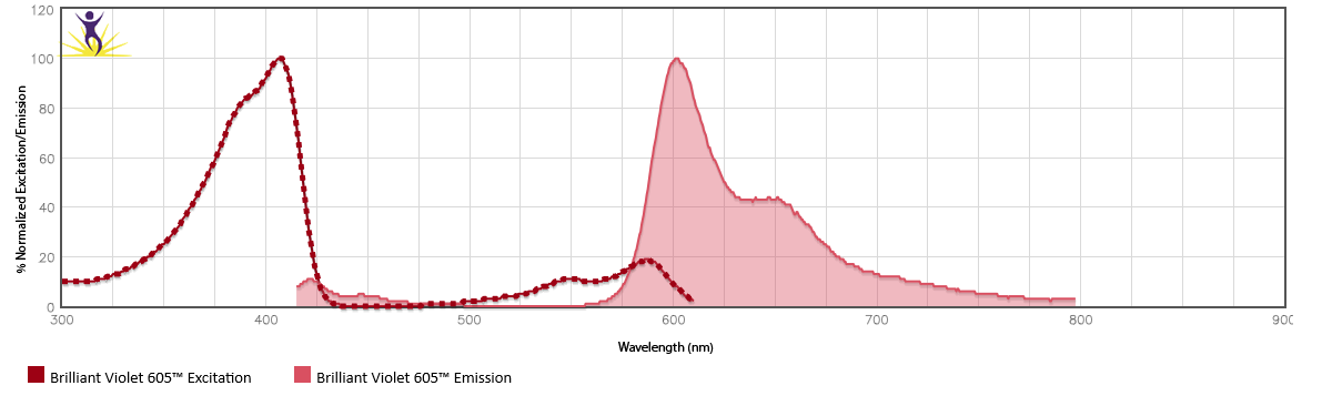
Brilliant Violet 605™ is a novel molecule based on the Brilliant Violet 421™ polymer core. It provides a much brighter alternative to eFluor® 605NC for multicolor flow on the violet laser and is a better alternative to nanocrystals for intracellular flow cytometry. Est. MW = 60 kD.
Excitation Max = 405 nm, Emission Max = 603 nm
Recommended filter = 610/20
Comparable Fluorophores: Qdot® 605, eFluor® 605NC
Brightness = 4 (On a scale from 1 to 5, with 5 being the brightest.)
Molar Extinction Coeff. = 2,400,000 M-1cm-1
Quantum Yield = 0.29 in DPBS
Beta Testing -
- T cell activation panel in rhesus macaques
- Comparison of cytokine expression on CD154+/CD8+ and CD154+/CD8- cells
Brilliant Violet 650™
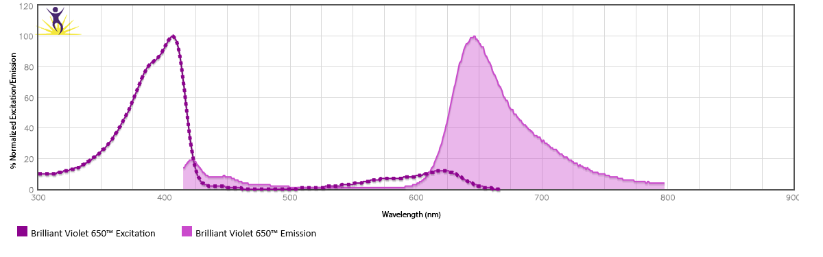
Brilliant Violet 650™ is a novel molecule based on the Brilliant Violet 421™ polymer core. It provides a much brighter alternative to eFluor® 650NC for multicolor flow on the violet laser and is a better alternative to nanocrystals for intracellular flow cytometry. Est. MW = 63 kD.
Excitation Max = 405 nm, Emission Max = 645 nm
Recommended filter = 660/20
Comparable Fluorophores: Qdot® 655, eFluor® 650NC
Brightness = 4 (On a scale from 1 to 5, with 5 being the brightest.)
Molar Extinction Coeff. = 2,500,000 M-1cm-1
Quantum Yield = 0.17 in DPBS
Beta Testing -
Brilliant Violet 711™
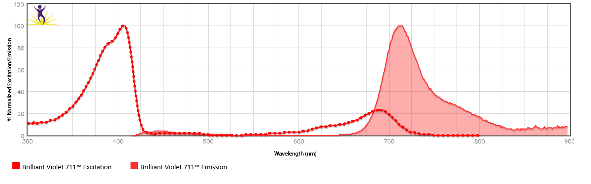
Brilliant Violet 711™ is a novel molecule based on the Brilliant Violet 421™ polymer core. It provides a much brighter alternative to eFluor® 700NC for multicolor flow on the violet laser and is a better alternative to nanocrystals for intracellular flow cytometry. Est. MW = 70 kD.
Excitation Max = 405 nm, Emission Max = 711 nm
Recommended filter = 710/50
Comparable Fluorophores: eFluor® 700NC, Qdot® 705
Brightness = 5 (On a scale from 1 to 5, with 5 being the brightest.)
Molar Extinction Coeff. = 2,800,000 M-1cm-1
Quantum Yield = 0.15 in DPBS
Beta Testing -
- Cytokine profiling of T cell subsets using Brilliant Violet™ fluorophores
- 15 Color Assay using Brilliant Violet™ Fluorophores
Brilliant Violet 750™
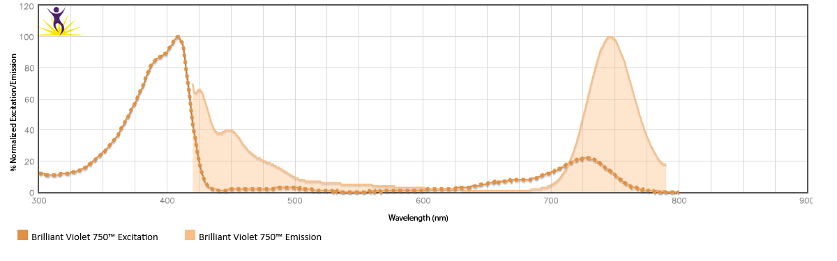
Brilliant Violet 750™ is a novel molecule based on the Brilliant Violet 421™ polymer core. It provides further options for the violet laser, particularly for those with either a spectral detection cytometer or a cytometer with a decagon configuration for the violet laser. Alternatively, it can be used in place of BV785™ on a standard violet laser octagon configuration. Est. MW = 70 kD.
Excitation Max = 405 nm, Emission Max = 750 nm
Recommended filter = 780/60 with a 740 LP
Comparable Fluorophores: None.
Brightness = 3 (On a scale from 1 to 5, with 5 being the brightest.)
Molar Extinction Coeff. = 2,301,131 M-1cm-1
Quantum Yield = 0.057 in DPBS
Brilliant Violet 785™
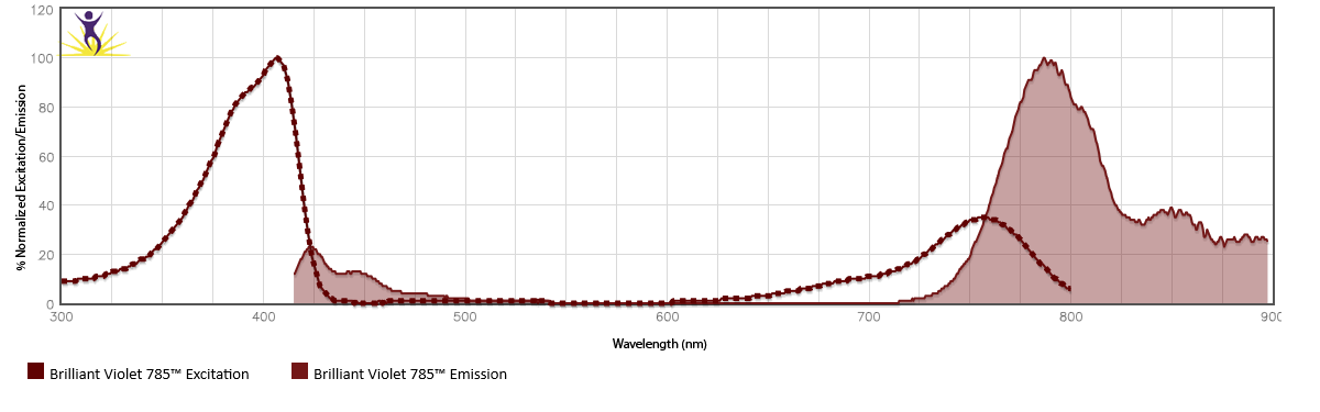
Brilliant Violet 785™ is a novel molecule based on the Brilliant Violet 421™ polymer core. It provides further options for the violet laser and is a better alternative to nanocrystals for intracellular flow cytometry. Est. MW = 60 kD.
Excitation Max = 405 nm, Emission Max = 785 nm
Recommended filter = 780/60
Comparable Fluorophores: QDot® 800
Brightness = 4 (On a scale from 1 to 5, with 5 being the brightest.)
Molar Extinction Coeff. = 2,500,000 M-1cm-1
Quantum Yield = 0.04 in DPBS
Beta Testing -
Learn the answers to Frequently Asked Questions about Brilliant Violet™ conjugates…
Brilliant Technology
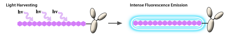
The Brilliant Violet™ family of fluorescent molecules are organic polymers with an extraordinary capacity to absorb energy (extinction coefficient) and a high efficiency with which to convert that absorbed energy to an emitted signal (quantum yield). When conjugated to antibodies, this results in high intensity brightness on labeled cells.
Improvements over Traditional Organic Dyes
Traditional organic dyes are quite small, ranging between 300 - 1200 Da. Although multiple dyes can be conjugated to a single antibody, each acts independently from the others around them, thus their potency is limited to their own structural constraints and the potential for self quenching. In contrast, the Brilliant Violet™ polymers are comparable in size to AmCyan or APC, but unlike a protein, consist of repeating fluorescent subunits that act cooperatively along the entire length of the polymer backbone. Energy is conducted much like a molecular antennae to capture light and pass it down the antennae like a lightning rod. As the subunits work cooperatively, the polymers' extinction coefficient is substantially larger than that of standard organic dyes and this is reflected in superior brightness and signal-to-noise.
Brilliant Violet™ Chemistry
Structurally, Brilliant Violet™ polymers are unsaturated organic materials comprised of alternating single and double bonds and aromatic units. It is this repeating bond structure that creates a continuous π-orbital system and extended electronic delocalization. Such features give rise to unique and tunable optical properties, including large extinction coefficients (>106 M-1cm-1), intense photoluminescence, and massive collective response, all of which help to address fundamental limitations in detection sensitivity. Adaptation of polymer side chain chemistry to impart solubility in aqueous solution confers ability for use as a highly sensitive fluorescent conjugate in biological applications such as flow cytometry and microscopy.
Physical properties, such as high quantum yield in typical flow buffers, high solubility, and minimal non-specfic binding, are all built into the backbone structure and in the side chain modifications. Additionally, the polymer design specifically incorporates well defined functional sites for covalent attachment to antibodies.
Brilliant Advantages
High Sensitivity Fluorescence™
Flourochromes derived from conducting polymers represent a new paradigm, High Sensitivity Fluorescence™, which aims to address fundamental shortcomings in reagents currently available for violet excitation in flow cytometry. Common dyes like Pacific Blue™, Alexa Fluor® 488 and Cy5 have high quantum yields but limited extinction coefficients. Alternative reporters like phycobiliproteins offer much greater absorbance cross-sections, producing brighter signals, but are limited by rapid photobleaching and sensitivity to fixation. Because the extinction coefficient of a conjugated polymer is directly proportional to the degree of polymerization (or number of repeat units), these structures are designed for improved brightness. Further, as these materials are derived from common synthetic organic and polymer chemistry techniques it is possible to manufacture reagents which are more defined and reproducible, in terms of size, conjugation sites, physical properties, and optical properties.
Unlike quantum dots, conjugated polymers have discrete excitation spectra, similar to that of organic dyes, which minimizes potential issues with cross-beam compensation.
Physical Properties
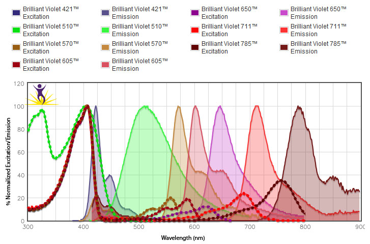
Brilliant Violet 421™ has an extinction coefficient of 2,500,000 M-1cm-1 at 405 nm, an aqueous solution quantum yield of 65 ± 5% and solubility in excess of 50 mg/mL in PBS. The extinction coefficient contributes to its superior brightness compared to Pacific Blue™, which has an extinction coefficient of 30,000 M-1cm-1. High Sensitivity Fluorescence™ polymers can also be modified with functional groups to produce high stokes shift emissions. Brilliant Violet 570™, Brilliant Violet 605™, and Brilliant Violet 650™ are such variants of the Brilliant Violet 421™ polymer, emitting maximally at 570 nm, 603 nm, and 645 nm, respectively. The figure below provides the excitation and emission spectra of the Brilliant Violet™ fluorophores. These can also be compared to other fluorophores in our Spectra Analyzer tool.
Brilliant Violet™antibody conjugates will change the future of flow cytometry, bringing new power to the violet laser. In particular, Brilliant Violet 421™ antibodies consistently stain at similar levels to PE, the brightest known fluorochrome, bringing unparalleled sensitivity and resolution to the violet laser, while Brilliant Violet 570™ antibodies add needed versatility to panel selection for multicolor flow cytometry. Brilliant Violet 605™ and Brilliant Violet 650™ antibodies also provide exceptionally bright signal and further expand options for multicolor panels. Brilliant Violet 711™ and Brilliant Violet 785™ bring newly expanded capability and selection for multicolor flow cytometry.

RBC-lysed human whole blood cells were stained with anti-CD3 conjugated to BV421™, PE, Pacific Blue™ or BD Horizon™ V450, and run on the BD™ LSR II flow cytometer. The stain index values indicated are derived at the optimal concentration for each conjugate. Stain index = (Median Fluorescence Intesity positive cells - Median Fluorescence Instensity negative cells)/2 x rSD.

RBC-lysed human whole blood cells were stained with anti-CD8 conjugated to BV570™, Pacific Orange™ or BD Horizon™ V500, and run on the BD™ LSR II flow cytometer. The stain index values indicated are derived at the optimal concentration for each conjugate.

Human whole blood cells were stained with anti-CD8 conjugated to BV605™, or eFluor® 605NC, and run on the BD™ LSR II flow cytometer. The stain index values indicated are derived at the optimal concentration for each conjugate.

Human whole blood cells were stained with anti-CD8 conjugated to BV650™, or eFluor® 650NC, and run on the BD™ LSR II flow cytometer. The stain index values indicated are derived at the optimal concentration for each conjugate.
Easy to Use and Trouble-Free
Brilliant Violet™ antibodies are simple to use, compatible with standard staining buffers, and stable to fixation. Provided in convenient 5 µl test sizes at optimal ready-to-use concentrations, our Brilliant Violet™ antibody products can easily be added to your multi-color panels. To compare fluorescence spectra data of Brilliant Violet™ conjugates with our other fluorochromes use the BioLegend Fluorescence Spectra Analyzer.
To help with flow panel construction using BioLegend’s Brilliant Violet™ antibodies, use our Multicolor Panel Selector web tool.
Extensive Antibody Selection
BioLegend provides an expansive selection of antibody specificities for Brilliant Violet 421™, Brilliant Violet 570™, Brilliant Violet 605™, Brilliant Violet 650™, Brilliant Violet 711™, and Brilliant Violet 785™. All products are manufactured by our expert chemists in San Diego, CA and are supported by our 100% satisfaction guarantee. For samples or custom conjugations, contact our sales team.

CD127
Human PBMCs were stained with anti-CD3 FITC and anti-CD127 (clone HCD127) conjugated to the BV421™ or Pacific Blue™. The data demonstrates the resolving power of BV421™, clearly showing separation of CD127-positive cells from dim and negative cells which can be difficult to define using Pacific Blue™

CD56
Human RBC-lysed whole blood cells were stained in an 11-color panel including HLA-DR PE/Cy7 and anti-CD56 (clone HCD56) conjugated to the BV421™ or anti-CD56 (clone MEM-188) Pacific Blue™. The data reveals discreet resolution of CD56 positive populations with BV421™. Data provided by Axel Schulz / Andreas Thiel, Charité - University Medicine Berlin.

Tetramer
Mouse spleen cells were labeled with PBS57-loaded mouse CD1d tetramer bound to Streptavidin-BV421™, -PE, or -Pacific Blue™. The data shows optimal staining with BV421™ when comparing equivalent tetramer concentration at 0.625 µg/ml. Data provided by Dr. Rick Willis, Emory/Yerkes. BioLegend does not currently offer catalog or custom CD1 tetramers.

Pooled Balb/c spleen and lymph node cells were stained with CD4-BV421™ or CD4-PB and sorted for CD4 positive cells. The cells were then labeled with CFSE and stimulated in 96-well flat-bottom plates coated with anti-CD3/anti-CD28 (1 µg/ml each) at 1 x 10⁵ cells/well. On day 4, cells were analyzed for CFSE dilution. Figure A demonstrates comparable division and expansion of cells sorted by BV421™ compared to those sorted by PB, as indicated by the loss of CFSE signal. Unstimulated cells are displayed as the gray histogram. Figure B shows the median fluorescence values of each group. Error bars represent the standard deviation of triplicate wells. Figure C demonstrates that the % of cells that have divided is comparable between BV421™ and PB-stained cells. Overall, the data shows viability of cells after staining and sorting with a BV421™ antibody. Data provided by Aras Toker/Jochen Hühn, Helmholtz Centre for Infection Research.

Human RBC-lysed whole blood cells were stained with (A) anti-CD8 BV570™ and anti-CD56 BV421™ or (B) anti-CD127 BV570™ and anti-CD25 BV421™. In B, cells were gated on CD4 positive cells. The data demonstrates the capabilities for using BV570™ and BV421™ conjugates together in multicolor panels. Data in B, provided by Eva Tolosa, University Medical Center Hamburg-Eppendorf.

PMA/ionomycin-stimulated (6 hours) human peripheral blood lymphocytes were surface stained with CD3 FITC and then intracellularly stained with IFN-γ (clone 4S.B3) Brilliant Violet 570™ (right) or mouse IgG1, k Brilliant Violet 570™ isotype control (left).


Here at BioLegend, we are impartial when it comes to instrumentation for flow cytometry. Our reagents work beautifully on instruments from all manufacturers for diverse biological applications. But as this field of single cell, fluorescence-based multiplexing continues to grow in total multicolor capacity, sensitivity and ease of use, we can’t wait to release reagents to help match pace with these innovative instruments.
The Cytek Aurora™ Spectral Cytometer is one of these instruments. The introductory configuration of this instrument is a 3-laser system (405 nm, 488 nm and 633 nm) and provides the possibility for 48 different fluorescent parameters with 2 scatter channels. This is only the theoretical limit as we continue to strive to provide fluorophores that are distinct enough to be accurately unmixed on the system. The Aurora™ also utilizes APD (Avalanche Photodiode) detectors, which liberate the near-IR and infrared wavelengths for more efficient detection of emitted photons in this range.
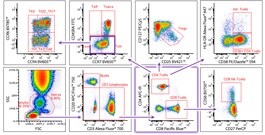
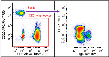
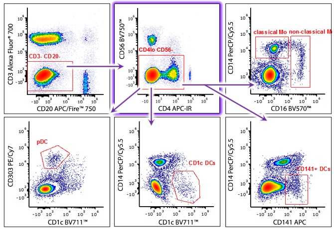
We will expand on data generated in-house here at BioLegend in this section as we test the limits of spectral flow cytometry, but in the meantime, here is a glimpse of 21 color data from 3 lasers with the use of fluorophores that spectrally overlap almost identically on traditional machines. For example, in the panel to the right, PerCP and PerCP/Cy5.5, Alexa Fluor® 647 and APC, Brilliant Violet 421™ and Pacific Blue™ are all utilized at the same time. It also allows researchers to use eight Brilliant Violet™ (BV421™, BV510™, BV570™, BV605™, BV650™, BV711™, the new BV750™, and BV785™) fluorophores together with minimal compensation concerns. If you have additional questions on this panel, feel free to contact technical service.
(Right) Human whole blood was stained with 21 different antibody conjugates and analyzed on the Cytek Aurora™ Spectral Flow Cytometer.
| Specificity | Format |
|---|---|
| CD141 | APC |
| CD303 | PE/Cy7 |
| CD16 | BV570™ |
| CD14 | PerCP/Cy5.5 |
| CLEC9A | PE |
| CD1c | BV711™ |
| IgD | BV510™ |
| CD20 | APC/Fire™ 750 |
| CD4 | APC-IR |
| CD8 | Pacific Blue™ |
| CD3 | Alex Fluor® 700 |
| CD25 | BV421™ |
| CCR6 | BV785™ |
| CD56 | BV750™ |
| CD45RA | FITC |
| CD27 | PerCP |
| CD127 | PE/Cy5 |
| CD38 | PE/Dazzle™ 594 |
| HLA-DR | Alexa Fluor® 647 |
| CD197 | BV650™ |
| CCR4 | BV605™ |
Brilliant Microscopy
Where BD Horizon™ V450, Pacific Blue™ and eFluor® 405 are just too dim for fluorescence microscopy, Brilliant Violet 421™ now enables visualization of antigens with directly conjugated antibodies, enhancing your capabilities with multi-color microscopy.
Historically, the "blue" channel on a microscope is reserved for an Hoechst or DAPI counterstaining due to the dim signal of typical dyes in that range, such as Alexa Fluor® 350, coumarin and Pacific Blue™ and the high sample autofluorescence often seen at that short wavelength of excitation. The following figures demonstrate that even on antibodies against markers that are relatively low in abundance, the Brilliant Violet 421™ direct conjugates can be used in imaging applications with high sensitivity. Further, they demonstrate extremely good photostability for repeated imaging and confocal z-stack capture. Also, these direct conjugates of Brilliant Violet 421™ demonstrate that there is no need for secondary antibodies or other amplificatiton techniques in order to achieve sufficient signal. Brilliant Violet 421™ will enable the addition of another color to a multicolor microscopy application on instruments capable of exciting this fluorophore between 360-420 nm.

Figure A. Photostability Curves plotting Brilliant Violet 421™ with and without antifade in the mounting medium against Pacific Blue™ mounted with no antifade. Photobleaching was conducted on a 3i spinning disk confocal, laser power at 100%, 300ms exposures every 1 s for a duration of 130 seconds. The curve is plotted as percent fluorescence relative to the initial fluorescence intensity. Since data is normalized to 100%, it is not a reflection on the initial fluorescence intensity for each fluorophore as BV421™ is much brighter than Pacific Blue™, as demonstrated in Figure B. With or without antifade, BV421™ retains more than half-maximal signal even after 130 seconds. Pacific Blue™, on the other hand, loses fluorescence more quickly, showing only 50% intensity by 60 seconds. Prolong Gold was able to attenuate the loss of fluorescence intensity for BV421™ upon initial exposure to laser, particularly noticeable within the first 10 to 30 seconds. Since most images are typically acquired within that initial few seconds, it is recommended to use a mounting medium containing antifade to preserve the brightest signal.
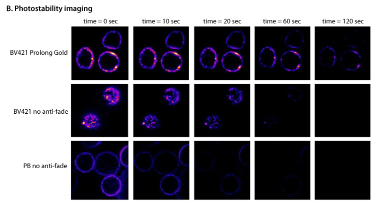
Figure B. Still images were captured at 0, 10, 20, 60, and 120 seconds. Samples were human PBMCs labeled with Brilliant Violet 421™ CD3 or Pacific Blue™ CD45. CD45 was required for detection of Pacific Blue™ because of its higher abundance than CD3 on PBMCs. Even though Pacific Blue™ endured 60 seconds of exposure before it lost 50% of its intensity, it was no longer bright enough at 20 seconds to be useful in imaging. The Brilliant Violet™ conjugates, on the other hand, were extremely bright and stable with antifade. Even at 120 seconds, a useful image could still be captured when antifade was used.
Brilliant Imaging
For multi-color imaging, the use of directly conjugated antibodies is essential. Brilliant Violet™ brings new capabilities to the violet laser, allowing for bright staining of weakly expressed markers.

NK92 cells (human NK cell line) were fixed and stained with anti-CD56 BV421, anti-perforin FITC, and phalloidin AF568 imaged on an Olympus IX81 spinning disk confocal microscope on 100X objective, NA 1.45. Exposures: 488 = 1000 ms, 568 = 100 ms, BV421™ (450 nm) = 200 ms. Data provided by Emily Mace and Jordan Orange, University of Pennsylvania.
Brilliant Violet 421™ Filter Choice
As previously mentioned, BV421™ is used in the “blue” channel, which is typically occupied by DAPI or Alexa Fluor® 405. However, as there is no set BV421™ filter on the market yet and because a “DAPI filter” may not be ideal, filter choice based on their wavelengths (excitation, emission and dichroic filters) is particularly important.
For example, the filter set in the image below works well as the dichroic filter (red line) does not block any of the BV421™ emission. However, in filter setups where this dichroic is set further to the right, a large percentage of the photons emitted by BV421™ may not reach the camera. If your filter setup is suitable, BV421™ can provide you with a bright, photostable option for multicolor microscopy.
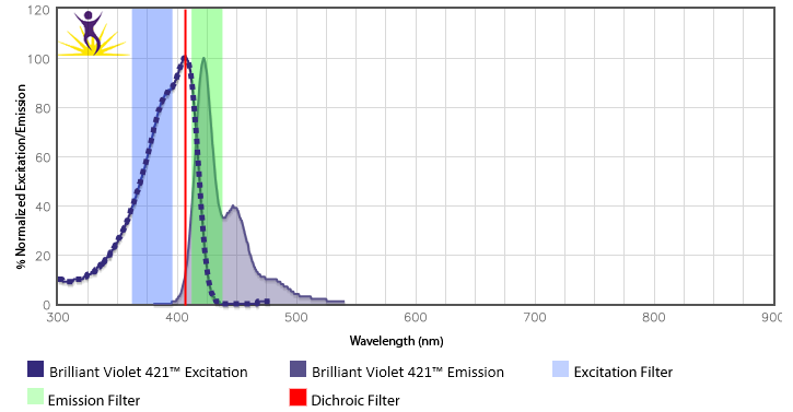
BioLegend has validated the following filter setups for BV421™ (Ex: Excitation, Em: Emission):
- Ex: 379/34, Dichroic: 409 LP, Em: 425/26
- Ex: 395/25, Dichroic: 405 LP, Em: 440/40
- Ex: 350/50, Dichroic: 400 LP, Em: 460/50
Brilliant High Throughput
BV421™ is useful for high thoughput content confocal imaging using the violet laser, providing clear and reliable results.

Primary human macrophages in 384-well plates were polarized to M1 or M2, then fixed and stained with Brilliant Violet 421™ CD38. Plate was read on the ImageXpress Ultra from Molecular Devices, and fluorescence signal was quantitated across 6 replicates. The mean fluorescence + SD is plotted. Data provided by unnamed collaborator.
Latest Publications
- Youssif C, et al. 2018. EMBO Mol Med 10:e8403. Pubmed
- Sharma PK, et al. 2018. Cancer Gene Ther 25:27. Pubmed
- Cho S, et al. 2018. Nat Commun 9:2757. Pubmed
- Zhang Y, et al. 2018. Nat Commun 9:6. Pubmed
- Goletz C, et al. 2018. Front Immunol 9:1614. Pubmed
- Frasca D, et al. 2018. PLoS One 13:e0197472. Pubmed
- Yarzabek B, et al. 2018. Elife 7:e34961. Pubmed
- Fernandez HR, et al. 2018. Cell Death Differ 25:1239. Pubmed
- Chattopadhyay A, et al. 2018. Sci Rep 8:9032. Pubmed
- Comoglio F, et al. 2018. Genome Res 28:295. Pubmed
- Young JS, et al. 2018. Front Immunol 9:1385. Pubmed
- García Nores GD, et al. 2018. Nat Commun 9:1970. Pubmed
- Ruscher R, et al. 2018. Bio Protoc 8:e2757. Pubmed
- Lee CH, et al. 2018. Elife 7:e32532. Pubmed
- Cobbold SP, et al. 2018. Front Immunol 9:1381. Pubmed
- Philippeos C, et al. 2018. J Invest Dermatol 138:811. Pubmed
- Asada S, et al. 2018. Nat Commun 9:2733. Pubmed
- Muir L, et al. 2018. Wellcome Open Res 2:97. Pubmed
- Mendt M, et al. 2018. JCI Insight 3. Pubmed
- Colacino JA, et al. 2018. Stem Cell Reports 10:1596. Pubmed
- Chung L, et al. 2018. Cell Host Microbe 23:203. Pubmed
- Liu S, et al. 2018. Front Immunol 9:1085. Pubmed
- Moore MJ, et al. 2018. Elife 7:e33057. Pubmed
- Dutton EE, et al. 2018. Wellcome Open Res 2:117. Pubmed
- Watanabe K, et al. 2018. JCI Insight 3. Pubmed
- Halim TYF, et al. 2018. Immunity 48:1195. Pubmed
- Echevarría-Vargas IM, et al. 2018. EMBO Mol Med 10:e8446. Pubmed
- Revathikumar P, et al. 2018. PLoS One 13:e0193210. Pubmed
- Friedensohn S, et al. 2018. Front Immunol 9:1401. Pubmed
- Lorentsen KJ, et al. 2018. Nat Commun 9:1679. Pubmed
- Böttcher JP, et al. 2018. Cell 172:1022. Pubmed
- Zukauskas A, et al. 2018. mSphere 3:e00303. Pubmed
- Geng X, et al. 2018. Aging (Albany NY) 10:1415. Pubmed
- Skucha A, et al. 2018. Nat Commun 9:1983. Pubmed
- Lyons J, et al. 2018. PLoS Biol 16:e2002417. Pubmed
- Hirota K, et al. 2018. Immunity 48:1220. Pubmed
- Rhys HI, et al. 2018. EBioMedicine 29:60. Pubmed
- Emmerson A, et al. 2018. J Clin Invest 128:3088. Pubmed
- Xu C, et al. 2018. Nat Commun 9:2449. Pubmed
- Kramann R, et al. 2018. JCI Insight 3. Pubmed
- Chen EW, et al. 2018. Sci Rep 8:10046. Pubmed
- Hsu JM, et al. 2018. Nat Commun 9:1908. Pubmed
- Compeer EB, et al. 2018. Nat Commun 9:1597. Pubmed
- Klepsch V, et al. 2018. Nat Commun 9:1538. Pubmed
- Wunderlich CM, et al. 2018. Nat Commun 9:1646. Pubmed
- Webster P, et al. 2018. Nat Commun 9:2649. Pubmed
- Espinosa JR, et al. 2018. Front Immunol 9:1371. Pubmed
- Van Dis E, et al. 2018. Cell Rep 23:1435. Pubmed
- Yamamoto R, et al. 2018. Cell Stem Cell 22:600. Pubmed
- Bellelli R, et al. 2018. Mol Cell 70:707. Pubmed
- Hossain DMS, et al. 2018. J Clin Invest 128:644. Pubmed
- Emgård J, et al. 2018. Immunity 48:120. Pubmed
- Bowers E, et al. 2018. Nat Med 24:95. Pubmed
- Gell JJ, et al. 2018. Stem Cell Res 27:46. Pubmed
- Felices M, et al. 2018. JCI Insight 3. Pubmed
- Kagoya Y, et al. 2018. Nat Commun 9:1915. Pubmed
- Carisey AF, et al. 2018. Curr Biol 28:489. Pubmed
- Watanabe J, et al. 2018. Oncotarget 9:24000. Pubmed
- Medvec AR, et al. 2018. Mol Ther Methods Clin Dev 8:65. Pubmed
- Ellis-Connell AL, et al. 2018. J Virol 92:e01748. Pubmed
- Garfall AL, et al. 2018. JCI Insight 3. Pubmed
- Yan Y, et al. 2018. JCI Insight 3. Pubmed
- Ferrando-Martinez S, et al. 2018. J Clin Invest 128:2089. Pubmed
- Lee SWL, et al. 2018. Front Immunol 9:416. Pubmed
- Idorn M, et al. 2018. Oncoimmunology 7:e1412029. Pubmed
- Setliff I, et al. 2018. Cell Host Microbe 23:845. Pubmed
- Maehara T, et al. 2018. Life Sci Alliance 1:e201800050. Pubmed
- Upadhyay AA, et al. 2018. Genome Med 10:20. Pubmed
- Warthan MD, et al. 2018. Biol Reprod 98:309. Pubmed
- Yang LH, et al. 2018. Cancer Manag Res 10:873. Pubmed
- Neff CP, et al. 2018. EBioMedicine 30:192. Pubmed
- Arce Vargas F, et al. 2018. Cancer Cell 33:649. Pubmed
- Verboven K, et al. 2018. Sci Rep 8:4677. Pubmed
- Floros VI, et al. 2018. Nat Cell Biol 20:144. Pubmed
- Sayin I, et al. 2018. J Exp Med 7:40286. Pubmed
- Peruzzotti-Jametti L, et al. 2018. Cell Stem Cell 22:355. Pubmed
- Li H, et al. 2018. PLoS Genet 14:e1007163. Pubmed
- Guedan S, et al. 2018. JCI Insight 3. Pubmed
- Thion MS, et al. 2018. Cell 172:500. Pubmed
- Jackson JT, et al. 2018. Blood Adv 2:347. Pubmed
- Toomey CB, et al. 2018. Invest Ophthalmol Vis Sci 59:662. Pubmed
- Duarte D, et al. 2018. Cell Stem Cell 22:64. Pubmed
- Catakovic K, et al. 2018. Oncoimmunology 7:e1371399. Pubmed
- BÖiers C, et al. 2018. Dev Cell 44:362. Pubmed
- Crncec I, et al. 2018. Mol Oncol 12:514. Pubmed
- LindenstrØm T, et al. 2018. EBioMedicine 27:27. Pubmed
- Howson LJ, et al. 2018. Nat Commun 9:253. Pubmed
- Bak RO, et al. 2018. Nat Protoc 13:358. Pubmed
- Garand M, et al. 2018. Front Immunol 9:257. Pubmed
- Natale CA, et al. 2018. Elife 7. Pubmed
- LaMothe RA, et al. 2018. Front Immunol 9:281. Pubmed
- Linde N, et al. 2018. Nat Commun 9:21. Pubmed
- Beraud C, et al. 2018. Virol J 15:35. Pubmed
- Guo B, et al. 2018. Nat Med 24:360. Pubmed
- Barnette DN, et al. 2018. JCI Insight 3. Pubmed
- Hsieh WC, et al. 2018. Nat Commun 9:463. Pubmed
- Efremova M, et al. 2018. Nat Commun 9:32. Pubmed
- Oyarce C, et al. 2018. Front Immunol 8:1794. Pubmed
- Zhang J, et al. 2018. Sci Rep 8:2373. Pubmed
- Pilbeam K, et al. 2018. Oncotarget 9:11938. Pubmed
- Martin SD, et al. 2018. Oncoimmunology 7:e1371895. Pubmed
- Lichtenegger FS, et al. 2018. Front Immunol 9:385. Pubmed
- Cao J, et al. 2018. Nat Commun 9:77. Pubmed
- Tran C, et al. 2017. J Immunol 10.4049/jimmunol.1600396. Pubmed
- Place D, et al. 2017. PLoS One 10.1371/journal.pone.0190384. Pubmed
- Koday M, et al. 2017. PLoS One 10.1371/journal.pone.0189780. Pubmed
- Reismann D, et al. 2017. Nat Commun. 10.1038/s41467-017-01538-9. Pubmed
- Petursdottir D, et al. 2017. Front Immunol. 10.3389/fimmu.2017.01699. Pubmed
- Chmielewski M and Abken H 2017. Cell Rep. 10.1016/j.celrep.2017.11.063. Pubmed
- Lecciso M, et al. 2017. Front Immunol. 10.3389/fimmu.2017.01918. Pubmed
- Hammer A, et al. 2017. Front Immunol. 10.3389/fimmu.2017.01922. Pubmed
- Castiblanco D, et al. 2017. Front Immunol. 10.3389/fimmu.2017.01833. Pubmed
- Mukhopadhyay M, et al. 2017. J Immunol 10.4049/jimmunol.1700953. Pubmed
- Vargas‐Inchaustegui D, et al. 2017. J Immunol 10.4049/jimmunol.1700586. Pubmed
- Pavelko K, et al. 2017. Front Immunol. 10.3389/fimmu.2017.01532. Pubmed
- Schuler F, et al. 2017. Nat Commun. 10.1038/s41467‐017‐01850‐4. Pubmed
- Scally S, et al. 2017. Nat Commun. 10.1038/s41467‐017‐01924‐3. Pubmed
- Maschmeyer P, et al. 2017. J Autoimmun. 10.1016/j.jaut.2017.11.005. Pubmed
- Palazon A, et al. 2017. Cancer Cell. 10.1016/j.ccell.2017.10.003. Pubmed
- Wright A, et al. 2017. J Allergy Clin Immunol. 10.1016/j.jaci.2017.04.025. Pubmed
- Day C, et al. 2017. J Immunol 10.4049/jimmunol.1700558. Pubmed
- Jackson E, et al. 2017. PLoS One 10.1371/journal.pone.0185160. Pubmed
- Wiese A, et al. 2017. PLoS One 10.1371/journal.pone.0184956. Pubmed
- Angkasekwinai P, et al. 2017. PLoS One 10.1371/journal.pone.0184684. Pubmed
- Baguma R, et al. 2017. PLoS One 10.1371/journal.pone.0184563. Pubmed
- Zheng Z, et al. 2017. PLoS One 10.1371/journal.pone.0184127. Pubmed
- Robert R, et al. 2017. PLoS One 10.1371/journal.pone.0184278. Pubmed
- Khare P, et al. 2017. J Autoimmun 10.1016/j.jaut.2017.09.002. Pubmed
- Bankoti R, et al. 2017. Sci Rep 10.1038/s41598-017-12171-3. Pubmed
- Trabanelli S, et al. 2017. Nat Commun 10.1038/s41467-017-00678-2. Pubmed
- Soukup K, et al. 2017. Sci Rep 10.1038/s41598-017-12208-7. Pubmed
- Jagannathan P, et al. 2017. Sci Rep 10.1038/s41598-017-10624-3. Pubmed
- DiPiazza A, et al. 2017. Sci Rep 10.1038/s41598-017-11313-x. Pubmed
- Dillinger B, et al. 2017. Front Immunol 10.3389/fimmu.2017.01152. Pubmed
- Bakke S,et al. 2017. J Immunol 10.4049/jimmunol.1700302. Pubmed
- Hamilton J,et al. 2017. J Immunol 10.4049/jimmunol.1700888. Pubmed
- Kamekura R,et al. 2017. J Immunol 10.4049/jimmunol.1601507. Pubmed
- Blanchfield L,et al. 2017. J Immunol 10.4049/jimmunol.1700792. Pubmed
- Keustermans G,et al. 2017. PLoS One 10.1371/journal.pone.0187068. Pubmed
- Gordon-Alonso M,et al. 2017. Nat. Commun 10.1038/s41467-017-00925-6. Pubmed
- Meng Y,et al. 2017. Cell Death Dis. 10.1038/cddis.2017.505. Pubmed
- Ayaub E,et al. 2017. Sci Rep. 10.1038/s41598-017-13511-z. Pubmed
- Carnevale G,et al. 2017. Sci Rep. 10.1038/s41598-017-14838-3. Pubmed
- Feng J,et al. 2017. Nat Commun. 10.1038/s41467-017-01056-8. Pubmed
- Moore T,et al. 2017. Cancer Immunol Immunother. 10.1007/s00262-017-2073-0. Pubmed
- Anstine L,et al. 2017. J Am Heart Assoc. 10.1161/JAHA.117.007097. Pubmed
- An X, et al. 2017. PLoS One. 10.1371/journal.pone.0181904. Pubmed
- Sia J, et al. 2017. PLoS Pathog. 10.1371/journal.ppat.1006530. Pubmed
- McCracken M, et al. 2017. PLoS Pathog. 10.1371/journal.ppat.1006487. Pubmed
- Svensson A, et al. 2017. PLoS One. 10.1371/journal.pone.0183268. Pubmed
- Huang L, et al. 2017. PLoS Biol. 10.1371/journal.pbio.2001750. Pubmed
- Martrus G, et al. 2017. PLoS One. 10.1371/journal.pone.0182532. Pubmed
- Mifuji K, et al. 2017. Bone Joint Res 10.1302/2046-3758.68.BJR-2016-0338.R1. Pubmed
- Bruder J, et al. 2017. Mol Ther Methods Clin Dev 10.1016/j.omtm.2017.08.003. Pubmed
- Poon E, et al. 2017. J Immunother Cancer 10.1186/s40425-017-0268-8. Pubmed
- Riou C, et al. 2017. Front Immunol 10.3389/fimmu.2017.00968. Pubmed
- Amelio P, et al. 2017. PLoS Negl Trop Dis 10.1371/journal.pntd.0005817. Pubmed
- Ries M, et al. 2017. PLoS Pathog 10.1371/journal.ppat.1006506. Pubmed
- Balakrishnan A, et al. 2017. Biol Blood Marrow Transplant 10.1016/j.bbmt.2017.07.016. Pubmed
- Johnson L, Olson B, and McNeel D. 2017. J Immunother Cancer 10.1186/s40425-017-0260-3. Pubmed
- Yao W, et al. 2017. EBioMedicine 10.1016/j.ebiom.2017.07.014. Pubmed
- Agelidis A, et al. 2017. Cell Rep 10.1016/j.celrep.2017.06.041. Pubmed
- Giudice V, et al. 2017. Cytometry A 10.1002/cyto.a.23162. Pubmed
- Liu Y, et al. 2017. Oncogene 10.1038/onc.2017.209. Pubmed
- Limbach K, et al. 2017. Malar J 10.1186/s12936-017-1911-z. Pubmed
- Szulc-D&acedil;browska L, et al. 2017. PLoS One 12(6):e0179166. Pubmed
- Bolton D, et al. 2017. PLoS Pathogens 13(6):e1006445. Pubmed
- Buschor S, et al. 2017. PLoS Pathogens 13(6):e1006476. Pubmed
- Pardo E, et al. 2017. PLoS One 12(6):e0177472. Pubmed
- Nenasheva T, et al. 2017. PLoS One 12(6):e0178983. Pubmed
- Joly P, et al. 2017. PLoS One 12(6):e0180568. Pubmed
- Lundtoft C, et al. 2017. PLoS Pathogens 13(6):e1006425. Pubmed
- Mitra T, et al. 2017. Vaccine 10.1016/j.vaccine.2017.06.035. Pubmed
- Gálvez-Cancino F, et al. 2017. Vaccine 10.1016/j.vaccine.2017.06.041. Pubmed
- Zheng G, et al. 2017. Respir Res 10.1186/s12931-017-0599-5. Pubmed
- Schofield L, et al. 2017. BMC Med 10.1186/s12916-017-0883-8. Pubmed
- Taraldsrud E, et al. 2017. J Autoimmun 10.1016/j.jaut.2017.04.004. Pubmed
- Kakizaki M, Watanabe R 2017. Neuropathology 10.1111/neup.12386. Pubmed
- Eddy W, et al. 2017. J Immunol 10.4049/jimmunol.1601777. Pubmed
- Nakachi S, et al. 2017. Arthritis Res Ther 10.1186/s13075-017-1309-x. Pubmed
- Shoda H, et al. 2017. Arthritis Res Ther 10.1186/s13075-017-1308-y. Pubmed
- Carbonetti S, et al. 2017. J Immunol Methods 10.1016/j.jim.2017.05.010. Pubmed
- Audigé A, et al. 2017. BMC Immunol 10.1186/s12865-017-0209-9. Pubmed
- Sordé L, et al. 2017. Imm Inflam Dis 10.1002/iid3.167. Pubmed
- Rutishauser L, et al. 2017. AIDS Res Hum Retroviruses 10.1089/AID.2016.0324. Pubmed
- Stratigou V, et al. 2017. Rheumatology 10.1093/rheumatology/kex078. Pubmed
- Parackova Z, et al. 2017. Immunol Lett 10.1016/j.imlet.2017.04.009. Pubmed
- Sckisel G, et al. 2017. J Immunother Cancer 10.1186/s40425-017-0235-4. Pubmed
- Frasca D, et al. 2017. Cell Immunol 10.1016/j.cellimm.2017.04.007. Pubmed
- Younan P, et al. 2017. MBio 10.1128/mBio.00226-17. Pubmed
- Noir S, et al. 2017. Nucleic Acids Res 10.1093/nar/gkx203. Pubmed
- Harvey R, et al. 2017. Endocrinology 10.1210/en.2016-1832. Pubmed
- Pinto-Cardoso S, et al. 2017. Sci Rep 10.1038/srep43741. Pubmed
- Ward E, Fu H, Marelli-Berg F 2017. Methods Mol Biol 10.1007/978-1-4939-6931-9_15. Pubmed
- Kristensen A, et al. 2017. Cytokine 10.1016/j.cyto.2017.02.017. Pubmed
- Nguyen T, et al. 2017. Clin Transl Immunology 10.1038/cti.2017.4. Pubmed
- Zhu X, et al. 2017. Arch Oral Biol 10.1016/j.archoralbio.2017.03.010. Pubmed
- Fisher J, et al. 2017. Mol Ther 10.1016/j.ymthe.2017.03.002. Pubmed
- Siedlik J, et al. 2017. J Immunol Methods 10.4049/jimmunol.1700003. Pubmed
- Yin Z, et al. 2017. Neurobiol Aging 10.1016/j.neurobiolaging.2017.03.021. Pubmed
- van der Velden V, et al. 2017. J Immunol Methods 10.1016/j.jim.2017.03.011. Pubmed
- Amara K, et al. 2017. J Autoimmun 10.1016/j.jaut.2017.03.004. Pubmed
- Eyquem J, et al. 2017. Nature 543:113-117. Pubmed
- Dotsey E, et al. 2017. Sci Rep 7:42584. Pubmed
- Mizuno T, et al. 2017. Sci Rep 7:42714. Pubmed
- Wouters K, et al. 2017. Sci Rep 7:42665. Pubmed
- Zamarin D, et al. 2017. Nat Commun 8:14340. Pubmed
- Lin J, et al. 2017. Sci Rep 7:41722. Pubmed
- Geetha H. Mylvaganam, Daniel Rios 2017. Proc Natl Acad Sci U S A 114(8):1976-1981. Pubmed
- Montané E, et al. 2017. PLoS One 12(2):e0171294. Pubmed
- Ender F, et al. 2017. PLoS One 12(2):e0172446. Pubmed
- Chierico L, et al. 2017. PLoS One 12(2):e0171815. Pubmed
- Cortez-Toledo O, et al. 2017. PLoS One 12(2):e0171268. Pubmed
- Nkwouano V, et al. 2017. PLoS One 12(2):e0171817. Pubmed
- Hayashi M, et al. 2017. EBioMedicine 15:127-136. Pubmed
- Suzuki T, et al. 2017. Cell Rep 18(8):2045-2057. Pubmed
- Smulski C, et al. 2017. Cell Rep 18(9):2189-2202. Pubmed
- S Woyciechowski, M Hofmann, H Pircher 2017. Eur J Immunol 47:244-250. Pubmed
- Gamal W, et al. 2017. Sci Rep 7:37541. Pubmed
- Walker-Sperling V, et al. 2017. EBioMedicine 10.1016/j.ebiom.2017.01.034. Pubmed
- Liddelow S, et al. 2017. Nature 541:481-487. Pubmed
- Rao S, et al. 2017. Cell 168(3):503-516.e12. Pubmed
- Gardner P, et al. 2017. Sci Rep 7:40830. Pubmed
- Peter Morawski, Chen-Feng Qi, Silvia Boll 2017. Sci Rep 7:40838. Pubmed
- Locke F, et al. 2017. Mol Ther 25(1):285-295. Pubmed
- Lentucci C, et al. 2017. J Biol Chem 292:2754-2772. Pubmed
- Jyh Liang Hor, William R. Heath, Scott N. Mueller 2017. Sci Rep 7:41091. Pubmed
- Tauriainen J, et al. 2017. Sci Rep 7:40354. Pubmed
- Liu J, et al. 2017. Mucosal Immunol 10.1038/mi.2016.139. Pubmed
- Greiner G, et al. 2017. Blood 129(3):371-382. Pubmed
- Laurent C, et al. 2017. Brain 140(Pt 1):184-200. Pubmed
- Cerina M, et al. 2017. Brain Behav Immun 59:103-117. Pubmed
- Schütz C, et al. 2017. Leukemia 10.1038/leu.2017.9. Pubmed
- AC Belkina, JE Snyder-Cappione 2017. Cytometry A 91:175-179. Pubmed
- Lundell A, et al. 2017. Sci Rep 7:39904. Pubmed
- Caggiati A, et al. 2017. Aesthet Surg J 10.1093/asj/sjw211. Pubmed
- Takeshita Y, et al. 2016. Neurol Neuroimmunol Neuroinflamm 4(1):e311. Pubmed
- Ling Y, et al. 2016. Pharmacol Res S1043-6618(16)31329-9. Pubmed
- Andresen V, et al. 2016. Cell Death Dis 7:e2497. Pubmed
- Aguilera T, et al. 2016. Nat Commun 7:13898. Pubmed
- Yasuda T, et al. 2016. PLoS One 11:e0167952. Pubmed
- Wahl S, et al. 2016. Nature 541:81-86. Pubmed
- Sharpe S, et al. 2016. Tuberculosis (Edinb) 101:174-190. Pubmed
- Ahmed R, et al. 2016. Cell Rep 17:2811-2818. Pubmed
- Swanson P, et al. 2016. PLoS Pathog 12:e1006022. Pubmed
- Blanc P, et al. 2016. Nat Commun 7:13600. Pubmed
- Lubaki N, et al. 2016. PLoS Pathog 12:e1006031. Pubmed
- Aliota M, et al. 2016. PLoS Negl Trop Dis 10:e0005168. Pubmed
- Clement M, et al. 2016. PLoS Pathog 12:e1006050. Pubmed
- Mehedi M, et al. 2016. PLoS Pathog 12:e1006062. Pubmed
- Muto M, et al. 2016. J Am Soc Nephrol 10.1681/ASN.2016050496. Pubmed
- Lee L, et al. 2016. PLoS One 11:e0167693. Pubmed
- Dekhtiarenko I, et al. 2016. PLoS Pathog 12:e1006072. Pubmed
- RY H, et al. 2016. Oncoimmunology 6:e1249561. Pubmed
- Pascual G, et al. 2016. Nature 541:41-45. Pubmed
- Hadadi E, et al. 2016. Sci Rep 6:39035. Pubmed
- Roberts E, et al. 2016. PLoS One 11:e0168488. Pubmed
- Eriksson E, et al. 2016. Gene Ther 10.1038/gt.2016.80. Pubmed
- Wei C, et al. 2016. Cell Death Dis 7:e2489. Pubmed
- Mier-Aguilar C, et al. 2016. PLoS One 11:e0168155. Pubmed
- Campa C, et al. 2016. Sci Signal 9(459):ra124. Pubmed
- Chandele A, et al. 2016. J Virol 90(24):11259-11278. Pubmed
- Kerstein A, et al. 2016. J Autoimmun S0896-8411(16)30186-X. Pubmed
- Woodham A, et al. 2016. Papillomavirus Res 2:21-30. Pubmed
- Valladao A, et al. 2016. J Immunol 197(12):4541-4551. Pubmed
- Tomić A, et al. 2016. PLoS Pathog 12:e1006015. Pubmed
- Rowan A, et al. 2016. PLoS Pathog 12:e1006030. Pubmed
- Madge P, et al. 2016. Sci Rep 6:36012. Pubmed
- Reinert L, et al. 2016. Nat Commun 7:13348. Pubmed
- Kiniry B, et al. 2016. Mucosal Immunol 10.1038/mi.2016.100. Pubmed
- Leslie G, et al. 2016. PLoS Pathog 12:e1005983. Pubmed
- Negri D, et al. 2016. Mol Ther 24:2021-2032. Pubmed
- Sabouri Z, et al. 2016. Nat Commun 7:13381. Pubmed
- Sebina I, et al. 2016. PLoS Pathog 12:e1005999. Pubmed
- Briceño O, et al. 2016. PLoS One 11:e0166496. Pubmed
- Angela M, et al. 2016. Nat Commun 7:13683. Pubmed
- E O’Koren, R Mathew, D Saban 2016. Sci Rep 6:20636. Pubmed
- Yoon Y, et al. 2016. Stem Cell Reports 7:840-853. Pubmed
- Ahadome S, et al. 2016. JCI Insight 1:e87012. Pubmed
- Moderzynski K, et al. 2016. PLoS Negl Trop Dis 10:e0005089. Pubmed
- van Gils M, et al. 2016. Nat Microbiol 2:16199. Pubmed
- Borducchi E, et al. 2016. Nature 540:284-287. Pubmed
- Mackroth M, et al. 2016. PLoS Pathog 12:e1005909. Pubmed
- Zhu L, et al. 2016. J Cell Sci 129: 4238 - 4251. Pubmed
- Montes de Oca M, et al. 2016. Cell Rep 17:399-412. Pubmed
- Inui M, et al. 2016. Int Immunol 10.1093/intimm/dxw044. Pubmed
- Kurowska-Stolarska M, et al. 2016. J Allergy Clin Immunol S0091-6749(16)31132-0. Pubmed
- Yu S, et al. 2016. Mol Ther 10.1038/mt.2016.175. Pubmed
- Sage P, et al. 2016. Nat Immunol 17:1436-1446. Pubmed
- Hombrink P, et al. 2016. Nat Immunol 17:1467-1478. Pubmed
- Isitman G, et al. 2016. PLoS One 11:e0164517. Pubmed
- Blom K, et al. 2016. J Immunol 197: 2762 - 2771. Pubmed
- Galle-Treger L, et al. 2016. Nat Commun 7:13202. Pubmed
- Serr I, et al. 2016. Proc Natl Acad Sci U S A 113: E6659 - E6668. Pubmed
- Pérol L, et al. 2016. Nat Commun 7:13027. Pubmed
- Geiger R, et al. 2016. Cell 167:829-842. Pubmed
- Martrus G, et al. 2016. J Virol 90: 9018 - 9028. Pubmed
- Talker S, et al. 2016. J Virol 90: 9364 - 9382. Pubmed
- Haynes A, et al. 2016. Physiol Rep 4: e12951. Pubmed
- Hu X, et al. 2016. Nat Commun 7:13095. Pubmed
- Martins R, et al. 2016. Nat Immunol 17:1361-1372. Pubmed
- Liyanage S, et al. 2016. Exp Eye Res 151:160-70. Pubmed
- Marquardt N, et al. 2016. J Immunol 197: 3069 - 3075. Pubmed
- Watson D, et al. 2016. Biomaterials 105:195-205. Pubmed
- Woda M, et al. 2016. J Infect Dis 214: 1001 - 1009. Pubmed
- Ng E, et al. 2016. Nat Biotechnol 34:1168-1179. Pubmed
- Scala M, et al. 2016. J Virol 90: 8563 - 8574. Pubmed
- Aalderen M, et al. 2016. PLoS Pathog 12:e1005903. Pubmed
- Pandya J, et al. 2016. J Leukoc Biol 100: 823 - 833. Pubmed
- Moriyama M, et al. 2016. PLoS One 11:e0164799. Pubmed
- Coursey T, et al. 2016. Mucosal Immunol 10.1038/mi.2016.83. Pubmed
- Rodriguez-Ruiz M, et al. 2016. Cancer Res 76: 5994 - 6005. Pubmed
- Payne K, et al. 2016. J Leukoc Biol 100: 625 - 635. Pubmed
- Gorman M, et al. 2016. J Virol 90: 8212 - 8225. Pubmed
- Mittal S, et al. 2016. Stem Cell Reports pii: S2213-6711(16)30187-4. Pubmed
- LM S, et al. 2016. Cell Rep 16(12): 3286-96. Pubmed
- An G, et al. 2016. Blood 128: 1590 - 1603. Pubmed
- Zhang B, et al. 2016. Mol Cell 63: 976-89. Pubmed
- Policicchio B, et al. 2016. PLoS Pathog 12: 1005879. Pubmed
- Lu X, et al. 2016. Nat Commun 7: 12719. Pubmed
- Vicetti Miguel R, et al. 2016. PLoS One 11: 0162445. Pubmed
- Paquin-Proulx D, et al. 2016. J Immunol 197: 1843 - 1851. Pubmed
- Beidaq A, et al. 2016. J Immunol 197: 1567 - 1576. Pubmed
- Loh L, et al. 2016. Proc Natl Acad Sci U S A 113: 10133 - 10138. Pubmed
- Burian A, et al. 2016. PLoS One 11: 0163297. Pubmed
- Schaper F, et al. 2016. Rheumatology 10.1093/rheumatology/kew324. Pubmed
- Cabrera-Perez J, et al. 2016. J Immunol 197: 1692 - 1698. Pubmed
- Kamp M, et al. 2016. PLoS One 11: 0163750. Pubmed
- Marco Barros R, et al. 2016. Cell 167: 203-218. Pubmed
- Li B, Schmidt N 2016. PLoS One 11: 0162427. Pubmed
- Vogel R, Erez A, Altan-Bonnet G 2016. Nat Commun 7: 12428. Pubmed
- Pachnio A, et al. 2016. PLoS Pathog 12: 1005832. Pubmed
- Roura S, et al. 2016. Lab Invest 10.1038/labinvest.2016.100. Pubmed
- Liu Q, et al. 2016. Cell Death Dis 1.93125. Pubmed
- Ghosh D, et al. 2016. J Immunol 197: 1788 - 1800. Pubmed
- Wong G, et al. 2016. J Immunol 197: 1642 - 1649. Pubmed
- Blom R, et al. 2016. PLoS One 11: 0163539. Pubmed
- Koay H, et al. 2016. Nat Immunol 10.1038/ni.3565. Pubmed
- Chai Y, et al. 2016. PLoS One 11: 0162853. Pubmed
- Gordon E, et al. 2016. Proc Natl Acad Sci U S A 113: 8765 - 8770. Pubmed
- Fukasawa K, et al. 2016. Sci Rep 6:30918. Pubmed
- Shi L, et al. 2016. Nat Commun 7:12335. Pubmed
- Ishimori A, et al. 2016. Allergol Int pii: S1323-8930(16)30105-8. Pubmed
- Huynh L, et al. 2016. Sci Rep 6:31959. Pubmed
- Uchtenhagen H, et al. 2016. Nat Commun 7:12614. Pubmed
- AR P, et al. 2016. Circ Res 118: 400-409. Pubmed
- Clutton G, et al. 2016. Sci Rep 6:30749. Pubmed
- Kritikou J, et al. 2016. Sci Rep 6:30636. Pubmed
- Schommers P, et al. 2016. J Virol 90: 7579 - 7586. Pubmed
- Pizzolla A, et al. 2016. PLoS One 11: 0160407. Pubmed
- Wallin J, et al. 2016. Nat Commun 7:12624. Pubmed
- Demers K, et al. 2016. PLoS Pathog 12: 1005805. Pubmed
- Dan J, et al. 2016. J Immunol 197: 983 - 993. Pubmed
- Im S, et al. 2016. Nature 537:417-421. Pubmed
- Sun K, et al. 2016. J Exp Med 213: 1851 - 1864. Pubmed
- Calascibetta F, et al. 2016. J Virol 90: 7541 - 7551. Pubmed
- Kostadinova E, et al. 2016. Sci Rep 6:30943. Pubmed
- Dibra D, et al. 2016. Clin Cancer Res 22: 3876 - 3883. Pubmed
- Yang E, et al. 2016. J Immunol 197: 934 - 941. Pubmed
- Cornelius C, et al. 2016. EBioMedicine pii: S2352-3964(16)30329-2. Pubmed
- Damgaard R, et al. 2016. Cell 166:1215-1230. Pubmed
- Khaitan A, et al. 2016. PLoS One 11: 0161786. Pubmed
- Kleiman E, et al. 2016. Proc Natl Acad Sci U S A 113: E3911 - E3920. Pubmed
- Ma C, et al. 2016. J Exp Med 213: 1589 - 1608. Pubmed
- Loukov D, et al. 2016. J Leukoc Biol 100: 121 - 129. Pubmed
- Rother M, et al. 2016. J Immunol 197: 441 - 448. Pubmed
- Vieyra-Garcia P, et al. 2016. Clin Cancer Res 22: 3328 - 3339. Pubmed
- Collette N, et al. 2016. Bone 88:20-30. Pubmed
- Swaims-Kohlmeier A, et al. 2016. J Immunol 197: 368 - 376. Pubmed
- Bhoj V, et al. 2016. Blood 128: 360 - 370. Pubmed
- Kaveh D, et al. 2016. Vaccine 34:4003-4011. Pubmed
- Swarnkar G, et al. 2016. Sci Rep 6:29896. Pubmed
- Barrett N, et al. 2016. Cell Rep 16: 1039-1054. Pubmed
- Nagafuchi Y, et al. 2016. Sci Rep 6:29338. Pubmed
- Mayo L, et al. 2016. Brain 139: 1939 - 1957. Pubmed
- Nilsson A, et al. 2016. PLoS One 11: 0158369. Pubmed
- Fromentin R, et al. 2016. PLoS Pathog 12: 1005761. Pubmed
- Prooyen N, et al. 2016. PLoS One 12: 1005749. Pubmed
- Ferretti A, et al. 2016. J Immunol 197: 470 - 479. Pubmed
- Campisi L, et al. 2016. Nat Immunol 10.1038/ni.3512. Pubmed
- Aryal B, et al. 2016. Nat Commun 7:12313. Pubmed
- Jonas B, et al. 2016. PLoS One 11: 0159189. Pubmed
- Arlehamn C, et al. 2016. PLoS Pathog 12: 1005760. Pubmed
- Cunningham C, et al. 2016. PLoS Pathog 12: 1005356. Pubmed
- Yasuoka T, et al. 2016. PLoS One 11: 0157395. Pubmed
- Crichton M, et al. 2016. Sci Rep 6:27217. Pubmed
- Kong Y, et al. 2016. Clin Cancer Res 22: 3057 - 3066. Pubmed
- Ishizaka A, et al. 2015. J Virol 90: 5665 - 5676. Pubmed
- Joosten S, et al. 2016. PLoS Pathog 12: 1005687. Pubmed
- Takizawa F, et al. 2016. J Immunol 196: 4522 - 4535. Pubmed
- Schmid M, et al. 2016. PLoS One 12: 1005676. Pubmed
- Nie M, et al. 2016. Cell Death Dis 7:e2261. Pubmed
- Lin D, et al. 2016. Cancer Res 76: 3179 - 3188. Pubmed
- AlHossiny M, et al. 2016. Cancer Res 76: 3376 - 3386. Pubmed
- Smedley J, et al. 2016. PLoS One 11: 0157535. Pubmed
- Kathania M, et al. 2016. Nat Immunol 10.1038/ni.3488. Pubmed
- Ding Z, Dahlin J 2016. Sci Rep 6:28290. Pubmed
- O’Connor K, et al. 2016. Genes Immun 10.1038/gene.2016.27. Pubmed
- Stadinski B, et al. 2016. Nat Immunol 10.1038/ni.3491. Pubmed
- Abdel-Mohsen M, et al. 2016. PLoS Pathog 12: 1005677. Pubmed
- Bruno L, et al. 2016. Immunol Cell Biol 10.1038/icb.2016.49. Pubmed
- Kariminia A, et al. 2016. Blood 127: 3082 - 3091. Pubmed
- Rout N, et al. 2016. PLoS One 11: 0157407. Pubmed
- Spanier J, et al. 2016. Nat Commun 7:11804. Pubmed
- Vasu S, et al. 2016. Blood 127: 2879 - 2889. Pubmed
- Ivarsson M, et al. 2016. Mucosal Immunol 10.1038/mi.2016.50. Pubmed
- Castro I, et al. 2016. J Virol 90: 5280 - 5291. Pubmed
- Schrøder M, et al. 2016. PLoS One 11: 0157387. Pubmed
- Gadd V, et al. 2016. PLoS One 11: 0157771. Pubmed
- Chalan P, et al. 2016. J Rheumatol 43: 1008 - 1016. Pubmed
- Qualai J, et al. 2016. PLoS One 11: 0156605. Pubmed
- Dudley D, et al. 2016. Nat Commun 7:12204. Pubmed
- Dou D, et al. 2016. Nat Cell Biol 18: 595-606. Pubmed
- Kar S, Colino J, Snapper C 2016. J Immunol 196: 4204 - 4213. Pubmed
- Alwis R, et al. 2016. J Virol 90: 4771 - 4779. Pubmed
- Denk F, et al. 2016. Cell Rep 15: 1771-1781. Pubmed
- Coppin E, et al. 2016. J Immunol 196: 4110 - 4121. Pubmed
- Wei H, et al. 2016. J Immunol 196: 3537 - 3541. Pubmed
- Mitchell E, et al. 2016. Nat Commun 7:11492. Pubmed
- Wheatley A, et al. 2016. Sci Rep 6: 26478. Pubmed
- D’Amico L, et al. 2016. J Exp Med 213: 827 - 840. Pubmed
- Stegmann K, et al. 2016. Sci Rep 6: 26157. Pubmed
- Heiden M, et al. 2016. Sci Rep 6: 26892. Pubmed
- Hensley-McBain T, et al. 2016. J Virol 90: 4981 - 4989. Pubmed
- Swanstrom A, et al. 2016. J Virol 90: 4966 - 4980. Pubmed
- Lombardi M, et al. 2016. J Leukoc Biol 99: 711 - 721. Pubmed
- Wan X, Thomas J, Unanue E 2016. J Exp Med 213: 967 - 978. Pubmed
- Li H, et al. 2016. J Immunol 196: 4064 - 4074. Pubmed
- Bähr A, et al. 2016. PLoS One 11: 0155676. Pubmed
- Grenga I, et al. 2016. Clin Transl Immunology 0.265972222. Pubmed
- Pennington L, et al. 2016. Nat Commun 7:11610. Pubmed
- Cao W, et al. 2016. Nat Commun 7:11687. Pubmed
- Gross C, et al. 2016. Proc Natl Acad Sci U S A 113: 2973 - 2982. Pubmed
- Saxena S, et al. 2016. Stem Cell Reports 6: 692-703. Pubmed
- Offersen R, et al. 2016. J Virol 90: 4441 - 4453. Pubmed
- Shin C, et al. 2016. Immunol Lett 172: 21-28. Pubmed
- Long C, et al. 2016. Toxicol Sci 10.1093/toxsci/kfw074. Pubmed
- Norton S, et al. 2016. Clin Transl Immunology 5: e76. Pubmed
- Kloepper J, et al. 2016. Proc Natl Acad Sci U S A 113: 4476-4481. Pubmed
- Peterson T, et al. 2016. Proc Natl Acad Sci U S A 113: 4470-4475. Pubmed
- Chowdhury A, et al. 2015. J Virol 89: 8677-8686. Pubmed
- Engel I, et al. 2016. Nat Immunol 10.1038/ni.3437. Pubmed
- Bal S, et al. 2016. Nat Immunol 10.1038/ni.3444. Pubmed
- McWilliam H, et al. 2016. Nat Immunol 17: 531-537. Pubmed
- Futrega K, Doran W 2016. Sci Rep 6: 23886. Pubmed
- Thoms J, et al. 2016. Mol Cell Biol 36: 1222-1236. Pubmed
- Prodinger J, et al. 2016. J Leukoc Biol 99: 583-594. Pubmed
- Chu C, et al. 2016. Dis Model Mech 9: 473-481. Pubmed
- Eller MA, et al. 2016. J Virol 90: 4005-4016. Pubmed
- Ermert D, et al. 2016. PLoS Pathog 11: 1005043. Pubmed
- Serr I, et al. 2016. Nat Commun 7:10991. Pubmed
- Xiong Y, et al. 2016. J Immunol 196: 2526 - 2540. Pubmed
- Ryan J, et al. 2016. Proc Natl Acad Sci U S A 113: 1286 - 1295. Pubmed
- Ravimohan S, et al. 2016. Clin Infect Dis 62: 795 - 803. Pubmed
- Croasdell A, et al. 2016. J Immunol 196: 2742 - 2752. Pubmed
- Furusawa J, et al. 2016. PLoS Pathog 12: 1005507. Pubmed
- Bennett M, et al. 2016. Proc Natl Acad Sci U S A 113: 1738 - 1746. Pubmed
- Woods T, et al. 2016. J Virol 90: 2783 - 2793. Pubmed
- Ward D, et al. 2016. Haematologica 101: 286 - 296. Pubmed
- Solaymani-Mohammadi S, et al. 2016. J Leukoc Biol 99: 475 - 482. Pubmed
- Souilhol C, et al. 2016. Nat Commun 7:10784. Pubmed
- Mchedlidze T, et al. 2016. Mucosal Immunol 10.1038/mi.2016.20. Pubmed
- Baglaenko Y, et al. 2016. PLoS One 11: 0150515. Pubmed
- Castro-Amarante M, et al. 2016. J Virol 90: 2195 - 2207. Pubmed
- Jürchott K, et al. 2016. PLoS One 11: 0150812. Pubmed
- Baerenwaldt A, et al. 2016. J Immunol 196: 2561 - 2571. Pubmed
- Thongthip S, et al. 2016. Genes Dev 30: 645 - 659. Pubmed
- Teixeira L, et al. 2016. Sci Rep 6:23475. Pubmed
- Manuzak J, et al. 2016. J Immunol 196: 2401 - 2409. Pubmed
- Sei J, et al. 2016. PLoS One 11: 0152192. Pubmed
- Kimura K, et al. 2016. Neurol Neuroimmunol Neuroinflamm 3: e210. Pubmed
- He Z, et al. 2016. Exp Hematol 44:161-165. Pubmed
- Wanke-Jellinek L, et al. 2016. J Leukoc Biol 99: 483 - 493. Pubmed
- Wieten R, et al. 2016. PLoS One 11: 0149871. Pubmed
- Pillet S, et al. 2016. Clin Immunol S1521-6616:30035-3. Pubmed
- Westernberg L, et al. 2016. J Allergy Clin Immunol S0091-6749(16)00083-X. Pubmed
- Bolton D, et al. 2016. J Virol 90: 1321 - 1332. Pubmed
- Xu Z, et al. 2016. Nat Commun 7:10728. Pubmed
- Zahr A, et al. 2016. Nat Commun 7:10363. Pubmed
- Björkander S, et al. 2016. Sci Rep 6:22083. Pubmed
- Däbritz J, et al. 2016. Sci Rep 6:20584. Pubmed
- Uddback I, et al. 2016. Sci Rep 6:20137. Pubmed
- Ferreira R, et al. 2016. J Infect Dis 213: 669 - 673. Pubmed
- Younis R, et al. 2016. J Immunol 196: 1419 - 1429. Pubmed
- Ioannidis L, et al. 2016. J Immunol 196: 1227 - 1238. Pubmed
- Nduom E, et al. 2016. Neuro Oncology 18: 195 - 205. Pubmed
- Björklund &, et al. 2016. Nat Immunol 17:451-460. Pubmed
- Cowan J, et al. 2016. Cell Rep 14:1041-1048. Pubmed
- Ryan E, et al. 2016. PLoS Pathog 12: 1005412. Pubmed
- Graav G, et al. 2016. PLoS One 11: 0148604. Pubmed
- Elayeb R, et al. 2016. Haematologica 101: 209 - 218. Pubmed
- Gros A, et al. 2016. Nat Med 10.1038/nm.4051. Pubmed
- Chew G, et al. 2016. PLoS Pathog 12: 1005349. Pubmed
- Cuenca M, et al. 2016. J Immunol 196: 726 - 737. Pubmed
- Bazett M, Haston M 2016. Sci Rep 6:19189. Pubmed
- Chen M, et al. 2016. J Immunol 194: 2607-15. Pubmed
- Wiesner D, et al. 2016. J Immunol 196: 365 - 374. Pubmed
- Tyagi N, et al. 2016. Cancer Lett 370: 260-267. Pubmed
- James E, et al. 2016. PLoS Pathog 12: 1005375. Pubmed
- Nicholas K, et al. 2015. Cytometry A 10.1002/cyto.a.22799. Pubmed
- Lakshminarayanan V, et al. 2016. PLoS One 11: 0145920. Pubmed
- Singhal P, et al. 2016. Proc Natl Acad Sci U S A 113: 122 - 127. Pubmed
- Yasuma K, et al. 2016. PLoS Pathog 12: 1005372. Pubmed
- Frei A, et al. 2016. Nat Methods 10.1038/nmeth.3742. Pubmed
- Glaser K, et al. 2016. PLoS One 11: 0146898. Pubmed
- Chiu Y, et al. 2016. Sci Rep 6:19227. Pubmed
- Verma S, et al. 2016. J Virol 90: 650 - 658. Pubmed
- Grove K, et al. 2016. PLoS One 11: 0145961. Pubmed
- Lieberman L, et al. 2016. Neurology 86: 375 - 381. Pubmed
- Akhmetzyanova I, et al. 2016. J Immunol 196: 484 - 492. Pubmed
- Montes de Oca M, et al. 2016. PLoS Pathog 12: 1005398. Pubmed
- Yabas M, et al. 2016. PLoS One 11: 0146774. Pubmed
- Prasad S, et al. 2015. PLoS One 10: 0145457. Pubmed
- Muppidi J, et al. 2015. J Exp Med 212: 2213 - 2222. Pubmed
- Hogan T, et al. 2015. Proc Natl Acad Sci U S A 112: 6917 - 6926. Pubmed
- Bending D, et al. 2015. J Immunol 195: 5616 - 5624. Pubmed
- Mian S, et al. 2015. Nat Commun 6: 10004. Pubmed
- Paris R, et al. 2015. PLoS One 10: 0144767. Pubmed
- Thommen D, et al. 2015. Cancer Immunol Res 3: 1344 - 1355. Pubmed
- Jablonski K, et al. 2015. PLoS One 10: 0145342. Pubmed
- Shi H, et al. 2015. Nat Immunol . Pubmed
- Salzberger W, et al. 2015. PLoS One 10: 0145324. Pubmed
- Evans C, et al. 2015. Haematologica 100: 1508 - 1516. Pubmed
- Carpio V, et al. 2015. PLoS One 10: 0144654. Pubmed
- Trivedi S, et al. 2015. Vaccine 33: 7315-7327. Pubmed
- Weiskopf D, et al. 2015. J Infect Dis 212: 1743 - 1751. Pubmed
- Karlsson H, et al. 2015. PLoS One 10: 0144787. Pubmed
- Holland C, et al. 2015. J Immunol 195: 5827 - 5836. Pubmed
- Schuldt N, et al. 2015. PLoS One 10: 0145762. Pubmed
- Lin H, et al. 2015. Mol Ther Methods Clin Dev 2: 15046. Pubmed
- Nowak A, et al. 2015. Ann Oncol 26: 2483 - 2490. Pubmed
- Martins M, et al. 2015. J Virol 89: 10802 - 10820. Pubmed
- Müller P, et al. 2015. Sci Transl Med 7: 315ra188. Pubmed
- Nastasi C, et al. 2015. Sci Rep 5:16148. Pubmed
- Autenrieth S, et al. 2015. Clin Transl Immunology 4:50. Pubmed
- Chukkapalli S, et al. 2015. PLoS One 10: 0143291. Pubmed
- Simonetta F, et al. 2015. J Immunol 195: 4712 - 4720. Pubmed
- Bialas K, et al. 2015. Proc Natl Acad Sci U S A 112: 13645 - 13650. Pubmed
- Mairhofer D, et al. 2015. J Invest Dermatol 135: 2785-93. Pubmed
- Ito F, et al. 2015. PLoS One 10: 0143370. Pubmed
- Mashiko S, et al. 2015. J Allergy Clin Immunol 136: 351-359. Pubmed
- Eberhardt K, et al. 2015. Clin Infect Dis 61: 1615 - 1623. Pubmed
- Pombo C, et al. 2015. J Infect Dis 212: 1376 - 1386. Pubmed
- Alves C, et al. 2015. PLoS One 10: 0142972. Pubmed
- Jones D, et al. 2015. J Immunol 195: 4753 - 4759. Pubmed
- Zhang Q, et al. 2015. Sci Rep 5:15948. Pubmed
- Polak R, et al. 2015. Blood 126: 2404 - 2414. Pubmed
- Cameron G, et al. 2015. J Immunol 195: 4604 - 4614. Pubmed
- Rios D, et al. 2015. Mucosal Immunol 101038/mi. Pubmed
- Onodera T, et al. 2015. Sci Rep 5:16801. Pubmed
- Yuzugullu H, et al. 2015. Nat Commun 6: 8501. Pubmed
- Prota G, et al. 2015. Vaccine 410: 01290-6. Pubmed
- Colineau L, et al. 2015. PLoS One 10: e0140978. Pubmed
- Zhang Y, et al. 2015. PLoS Pathog 11: e1005167. Pubmed
- Tsuzuki S, et al. 2015. Int Immunol 10.1093/intimm/dxv058. Pubmed
- Gordon E, et al. 2015. Proc Natl Acad Sci U S A 112: 13075 - 13080. Pubmed
- Rainho J, et al. 2015. J Virol 89: 10625 - 10636. Pubmed
- Romano A, et al. 2015. J Immunol 195: 3816 - 3827. Pubmed
- Voigt A, et al. 2015. Biol Open 10.1242/bio.013771. Pubmed
- Sharkey A, et al. 2015. J Immunol 195: 3026 - 3032. Pubmed
- Lisovsky I, et al. 2015. J Virol 89: 9909 - 9919. Pubmed
- Evans H, et al. 2015. J Immunol 195: 2973 - 2984. Pubmed
- Marquardt N, et al. 2015. J Immunol 195: 3262 - 3272. Pubmed
- Culina S, et al. 2015. Diabetes 64: 3532 - 3542. Pubmed
- Herrera R, et al. 2015. Infect Immun 83: 3771 - 3780.. Pubmed
- Chowdhury A, et al. 2015. J Immunol 195: 3237 - 3247. Pubmed
- Blackburn M, et al. 2015. J Immunol 195: 3227 - 3236. Pubmed
- James BR, et al. 2014. Cancer Immunol Immunother 63:1213-1227. Pubmed
- Bramwell K, et al. 2015. J Immunol 195: 1647 - 1656. Pubmed
- Richardson ET, et al. 2015. PLoS One 10: 1371. Pubmed
- Colliou N, et al. 2015. Toxins 7: 3805 - 3817. Pubmed
- Wu X, et al. 2015. J Biol Chem 290: 23135-23147. Pubmed
- Schippers A, et al. 2015. Mucosal Immunol 101038/mi201582. Pubmed
- Dokun A, et al. 2015. Am J Physiol Heart Circ Physiol 309: 790-803. Pubmed
- White A, et al. 2015. Clin Vaccine Immunol 22: 992-1003. Pubmed
- Adachi Y, et al. 2015. J Exp Med 212: 1709-1723. Pubmed
- Lammers K, et al. 2015. PLoS One 10: 0138338. Pubmed
- Calderon B, et al. 2015. J Exp Med 212: 1497-1512. Pubmed
- Wang C, et al. 2015. Sci Rep 5: 14124. Pubmed
- Donia M, et al. 2015. Cancer Res 75: 3747-3759. Pubmed
- Meunier I, et al. 2015. PLoS One 10: 0138055. Pubmed
- McCarthy N, et al. 2015. J Immunol 195: 2675-2682. Pubmed
- Porter D, et al. 2015. Sci Transl Med 7: 303ra139. Pubmed
- Mattiola I, et al. 2015. J Immunol 195: 2818-2828. Pubmed
- Ellis G, et al. 2015. EMBO Rep 16: 1203-1218. Pubmed
- Terawaki S, et al. 2015. J Cell Biol 210: 1133-1152. Pubmed
- Marshall N, et al. 2015. Toxicol Sci 147: 127-139. Pubmed
- Causi E, et al. 2015. PLoS One 10: 0136717. Pubmed
- Cardoso M, et al. 2015. Infect Immun 83: 3534-3544. Pubmed
- Lee C, et al. 2015. Nat Commun 6: 8477. Pubmed
- Xu Y, et al. 2015. J Vis Exp 99: 52863. Pubmed
- Quah B, et al. 2014. J Vis Exp 88: 51627. Pubmed
- Noto A, et al. 2013. J Vis Exp 82: 51105. Pubmed
- Luck H, et al. 2015. Cell Metab 21 527 . Pubmed
- AM C, et al. 2015. Proc Natl Acad Sci U S A 112 7557 . Pubmed
- Agerstam H, et al. 2015. Proc Natl Acad Sci U S A 112: 10786-10791. Pubmed
- Hammitzsch A, et al. 2015. Proc Natl Acad Sci U S A 112: 10768-10773. Pubmed
- Pablo-Bernal R, et al. 2015. J Gerontol A Biol Sci Med Sci 101093/gerona/glv121. Pubmed
- Levy N, et al. 2015. J Immunol 195: 1713-1722. Pubmed
- Lofano G, et al. 2015. J Immunol 195: 1617-1627. Pubmed
- Rider P, et al. 2015. J Immunol 195: 1705-1712. Pubmed
- Lainé A, et al. 2015. J Immunol 195: 1791-1803. Pubmed
- García-Arriaza J, et al. 2015. J Virol 89: 8525-8539. Pubmed
- Weiskopf D, et al. 2015. Proc Natl Acad Sci U S A 112: E4256-E4263. Pubmed
- Johnson S, et al. 2015. J Virol 89: 7494-7505. Pubmed
- Williams B, et al. 2015. Dis Model Mech 8: 805-815. Pubmed
- Boyle M, et al. 2015. J Infect Dis 212: 416-425. Pubmed
- Kugler J, et al. 2015. Toxicol Sci 146: 374-385. Pubmed
- Jimeno R, et al. 2015. J Leukoc Biol 98: 257-269. Pubmed
- Sanders K, et al. 2015. Cancer Immunol Res 3: 891-901. Pubmed
- Thiele K, et al. 2015. Am J Pathol Available online 5 August 2015. Pubmed
- Kullberg M, et al. 2015. Nanomedicine 11: 1355-1363. Pubmed
- Ray A, et al. 2015. PLoS One 10: 0137314. Pubmed
- Leeansyah E, et al. 2015. PLoS Pathog 11: 1005072. Pubmed
- Rosenheinrich M, et al. 2015. PLoS One 10: 0136290. Pubmed
- Chen Y, et al. 2015. PLoS One 10: 0135217. Pubmed
- Santra S, et al. 2015. PLoS Pathog 11: 1005042. Pubmed
- Ganti SN, et al. 2015. Sci Rep 5: 12255. Pubmed
- Sd W, et al. 2015. Sci Rep 5: 12270. Pubmed
- Li J, et al. 2015. Nat Commun 6: 7737. Pubmed
- Di Niro R, et al. 2015. Mucosal Immunol . Pubmed
- Granja T, et al. 2015. Thromb Res . Pubmed
- Taguchi K, et al. 2015. J Urol . Pubmed
- Ma C, et al. 2015. J Allergy Clin Immunol . Pubmed
- Lee J, et al. 2015. Clin Immunol 159: 37-46. Pubmed
- McClellan S, et al. 2015. Methods 82: 47-54. Pubmed
- Kolan S, et al. 2015. PLoS One 10: 0134113. Pubmed
- Boyle M, et al. 2015. PLoS Pathog 11: 1005041. Pubmed
- Cardoso F, et al. 2015. PLoS One 10: 0131904. Pubmed
- Sanjo H, et al. 2015. PLoS One 10: 0128761. Pubmed
- Kim P, et al. 2015. Infect Immun 83: 2992 - 3002. Pubmed
- Inui M, et al. 2015. Int Immunol 27: 345 - 355. Pubmed
- Boer M, et al. 2015. Clin Vaccine Immunol 22: 778 - 788. Pubmed
- Fuhrman C, et al. 2015. J Immunol 195: 145 - 155. Pubmed
- Kuebler P, et al. 2015. Proc Natl Acad Sci U S A 112: 8379 - 8384. Pubmed
- Herz J, et al. 2015. J Exp Med 212: 1153 - 1169. Pubmed
- Koyama M, et al. 2015. J Exp Med 212: 1303 - 1321. Pubmed
- Allman W, et al. 2015. Proc Natl Acad Sci U S A 112: 4094 - 4103. Pubmed
- Yamamoto T, et al. 2015. Sci Transl Med 7: 298ra120. Pubmed
- Motozono C, et al. 2015. J Biol Chem 290: 18924 - 18933. Pubmed
- Toomey C, et al. 2015. Proc Natl Acad Sci U S A 112:3040. Pubmed
- Phiwpan K, et al. 2015. PLoS One 10:128198. Pubmed
- Shinohara M 2015. J Immunol 194:5595. Pubmed
- Mondini M, et al. 2015. Mol Cancer Ther 14:1336. Pubmed
- Marks-Bluth J, et al. 2015. Mol Cell Biol 35:2165. Pubmed
- Sharma S, et al. 2015. J Immunol 194:5529. Pubmed
- Jacque E, et al. 2015. J Exp Med 212:883. Pubmed
- Severson J, et al. 2015. Cancer Immunol Res 3:620. Pubmed
- Opata M, et al. 2015. J Immunol 194:5346. Pubmed
- Ortiz A, et al. 2015. J Virol 89:5883. Pubmed
- Doni A, et al. 2015. J Exp Med 212:905. Pubmed
- Mawa P, et al. 2015. Phil Trans R Soc B 19:370. Pubmed
- Rao T, et al. 2015. Stem Cell Res 14:307. Pubmed
- Misra R, et al. 2015. Hum Immunol 76:329. Pubmed
- Berent-Maoz B, et al. 2015. PLoS One 10:125414. Pubmed
- Holm J, et al. 2015. PLoS One 10:125495. Pubmed
- Hamilton J, et al. 2015. J Immunol 194:5022. Pubmed
- Zhou J, et al. 2015. J Immunol 194:4688. Pubmed
- Aalderen M, et al. 2015. J Virol 89:5110. Pubmed
- Siegemund S, et al. 2015. PLoS One 10:124661. Pubmed
- Chukkapalli S, et al. 2015. Pathog Dis 73:9. Pubmed
- Tsai C, et al. 2015. J Immunol 194:3890. Pubmed
- Liu B, et al. 2015. J Immunol 194:3583. Pubmed
- Lisovsky I, et al. 2015. J Leukoc Biol 97:761. Pubmed
- Sakaguchi S, et al. 2015. J Leukoc Biol 97:635. Pubmed
- Claiborne D, et al. 2015. Proc Natl Acad Sci U S A 112:1480. Pubmed
- Lang P 2015. Int Immunol 27:253. Pubmed
- Hu-Lieskovan S, et al. 2015. Sci Transl Med 7:279. Pubmed
- Wiesner D, et al. 2015. PLoS Pathog 11:1004701. Pubmed
- Kim Y, et al. 2015. PLoS One 10:120294. Pubmed
- Liao X, et al. 2015. PLoS One 10:118176. Pubmed
- Meijgaarden K, et al. 2015. PLoS Pathog 11:1004671. Pubmed
- Krishnaswamy J, et al. 2015. Proc Natl Acad Sci U S A 112:3056. Pubmed
- Steindor M, et al. 2015. PLoS One 10:119737. Pubmed
- Marquardt N, et al. 2015. J Immunol 194:2467. Pubmed
- Watson N, et al. 2015. J Immunol 194:2796. Pubmed
- Montezuma-Rusca J, et al. 2015. J Immunol 194:2561. Pubmed
- Michelet X, et al. 2015. J Immunol 194:2079. Pubmed
- Herbst S, et al. 2015. EMBO Mol Med 30:240. Pubmed
- Lucas A, et al. 2015. PLoS One 10:117160. Pubmed
- Breton G, et al. 2015. J Exp Med 212:401. Pubmed
- Lee J, et al. 2015. J Exp Med 212:385. Pubmed
- Afshar M, et al. 2015. Alcohol 49:57. Pubmed
- Cabrera-Perez J, et al. 2015. J Immunol 194:1609. Pubmed
- Karsten C, et al. 2015. J Immunol 194:1841. Pubmed
- Wang T, et al. 2015. Proc Natl Acad Sci U S A 112:440. Pubmed
- Price P, et al. 2015. J Immunol 194:1164. Pubmed
- Ratliff M, et al. 2015. J Immunol 194:940. Pubmed
- Zinöcker S, et al. 2015. J Immunol 194:929. Pubmed
- Gerna G, et al. 2015. J Gen Virol 96:360. Pubmed
- Milne P, et al. 2015. Blood 125:470. Pubmed
- Singh K, et al. 2015. Sci Rep 14:7767. Pubmed
- Kivit S, et al. 2015. Clin Immunol 156:28. Pubmed
- Badarau A, et al. 2015. J Biol Chem 290:142. Pubmed
- Neumann B, et al. 2015. J Leukoc Biol 97:19. Pubmed
- Weed D, et al. 2015. Clin Cancer Res 21:39. Pubmed
- Schaik B, et al. 2015. J Virol 89:568. Pubmed
- Huang L, et al. 2015. J Immunol 194:283. Pubmed
- Woda M, Mathew A 2014. J Immunol Methods 416:167. Pubmed
- Robinson A, et al. 2014. Am J Physiol Gastrointest Liver Physiol 307:1115. Pubmed
- Blanco R, et al. 2014. Sci Signal 2:354. Pubmed
- Peters N, et al. 2014. PLoS Pathog 10:1004538. Pubmed
- Ballet R, et al. 2014. PLoS Pathog 10:1004550. Pubmed
- Textor A, et al. 2014. Cancer Res 74:6796. Pubmed
- Braun M, et al. 2014. PLoS Pathog 10:1004521. Pubmed
- Willmann K, et al. 2014. Nat Commun 5:5360. Pubmed
- Kennedy J, et al. 2014. J Exp Med 211:2519. Pubmed
- Mirsoian A, et al. 2014. J Exp Med 211:2373. Pubmed
- Prete G, et al. 2014. Antimicrob Agents Chemother 58:6790. Pubmed
- Poh C, et al. 2014. Infect Immun 82:4854. Pubmed
- Mylvaganam G, et al. 2014. J Immunol 193:4527. Pubmed
- Gutierrez D, et al. 2014. Diabetes 63:3827. Pubmed
- Purdy A, et al. 2014. J Immunol 193:4675. Pubmed
- Bertuzzi M, et al. 2014. PLoS Pathog 10:1004413. Pubmed
- Luetke-Eversloh M, et al. 2014. PLoS Pathog 10:1004441. Pubmed
- Freeman A, et al. 2014. PLoS One 9:110928. Pubmed
- Xing Y, et al. 2014. J Neurosci 34:14128. Pubmed
- Jansen D, et al. 2014. Rheumatology 54:728. Pubmed
- Ohue Y, et al. 2014. Clin Cancer Res 20:5052. Pubmed
- Herati R, et al. 2014. J Immunol 193:3528. Pubmed
- Royle C, et al. 2014. J Immunol 193:3538. Pubmed
- Brandau S, et al. 2014. PLoS One 9:106903. Pubmed
- Hu H, et al. 2014. Proc Natl Acad Sci U S A 111:13439. Pubmed
- Payne T, et al. 2014. J Virol 88:9514. Pubmed
- Bending D, et al. 2014. J Immunol 193:2699. Pubmed
- Arlehamn C, et al. 2014. J Immunol 193:2931. Pubmed
- Lucera M, et al. 2014. J Virol 88:10803. Pubmed
- Choo J, et al. 2014. J Virol 88:10613. Pubmed
- Requena P, et al. 2014. J Immunol 193:2971. Pubmed
- Guo H, et al. 2014. J Leukoc Biol 96:419. Pubmed
- Du J, et al. 2014. Cancer Immunol Res 2:878. Pubmed
- Huang W, et al. 2014. J Immunol 193:2267. Pubmed
- Ochiai S, et al. 2014. J Immunol 193:2504. Pubmed
- Schick V, et al. 2014. Gut 63:1469. Pubmed
- Chen Z, et al. 2014. Cancer Immunol Res 2:911. Pubmed
- Chen J, et al. 2014. Cell Res 24:1050. Pubmed
- Cowan J, et al. 2014. J Immunol 193:1204. Pubmed
- Taguchi K, et al. 2014. J Am Soc Nephrol 25:1680. Pubmed
- Radovanovic I, et al. 2014. J Immunol 193:1290. Pubmed
- Riou C, et al. 2014. PLoS One 9:102178. Pubmed
- Mise-Omata S, et al. 2014. Int Immunol 26:607. Pubmed
- Sedlak C, et al. 2014. Dev Comp Immunol 45:97. Pubmed
- Weijer M, et al. 2014. Nat Commun 5:3832. Pubmed
- Gang A, et al. 2014. Blood Cancer J 4:197. Pubmed
- Lundell A, et al. 2014. J Immunol 193:510. Pubmed
- Atefi M, et al. 2014. Clin Cancer Res 20:3446. Pubmed
- Boudreau J, et al. 2014. PLoS One 9:99543. Pubmed
- Moreno M, et al. 2014. J Neurosci 34:8175. Pubmed
- Friedman R, et al. 2014. Proc Natl Acad Sci U S A 111:9223. Pubmed
- Marandu T, et al. 2014. Exp Gerontol 54:101. Pubmed
- Neubert K, et al. 2014. J Immunol 192:5830. Pubmed
- Terszowski G, et al. 2014. J Immunol 192:5618. Pubmed
- Abreu C, et al. 2014. PLoS One 9:97257. Pubmed
- Weber G, et al. 2014. J Exp Med 211:2519. Pubmed
- Demberg T, et al. 2014. Clin Immunol 153:264. Pubmed
- Lu D, et al. 2014. Cell Immunol 152:77. Pubmed
- Fredriksson K, et al. 2014. J Immunol 192:4497. Pubmed
- Lindner J, et al. 2014. J Immunol 192:4883. Pubmed
- Buggert M, et al. 2014. J Immunol 192:4685. Pubmed
- Cartwright E, et al. 2014. J Immunol 192:4666. Pubmed
- Claudepierre M, et al. 2014. J Virol 88:5242. Pubmed
- Tabler C, et al. 2014. J Virol 88:4976. Pubmed
- Škrnjug I, et al. 2014. PLoS One 9:95728. Pubmed
- Higdon L, et al. 2014. Proc Natl Acad Sci U S A 111:5652. Pubmed
- Xu Y, et al. 2014. Proc Natl Acad Sci U S A 111:6371. Pubmed
- Bunn P, et al. 2014. J Immunol 192:3709. Pubmed
- Pérez-Quintero L, et al. 2014. J Exp Med 211:727. Pubmed
- Lyngaa R, et al. 2014. Clin Cancer Res 20:1768. Pubmed
- Stanisic D, et al. 2014. J Infect Dis 210:295. Pubmed
- Tan Y, et al. 2014. Cell Immunol 151:55. Pubmed
- Zenker S, et al. 2014. J Immunol 192:2830. Pubmed
- White A, et al. 2014. J Immunol 192:2659. Pubmed
- Kim P, et al. 2014. J Immunol 192:2622. Pubmed
- Cherukuri A, et al. 2014. J Am Soc Nephrol 7:1575. Pubmed
- Wu W, et al. 2014. Proc Natl Acad Sci U S A 111:4221. Pubmed
- Buggert M, et al. 2014. J Immunol 192:2099. Pubmed
- Bouwer A, et al. 2014. J Immunol 192:2514. Pubmed
- Tameris M, et al. 2014. PLoS One 9:87340. Pubmed
- Blankenhaus B, et al. 2014. PLoS Pathog 10:1003913. Pubmed
- Altin J, et al. 2014. Proc Natl Acad Sci U S A 111:2067. Pubmed
- Chirkova T, et al. 2013. J Virol 87:13466. Pubmed
- Liu X, et al. 2013. Diabetes 62:4228. Pubmed
- Bigley A, et al. 2013. Brain Behav Immun 889:1591. Pubmed
- Hardy C, et al. 2013. J Immunol 191:5278. Pubmed
- Scally S, et al. 2013. J Exp Med 210:2569. Pubmed
- Sugiyama D, et al. 2013. Proc Natl Acad Sci U S A 110:17945. Pubmed
- Johansson J, et al. 2013. J Neurosci 33:16016. Pubmed
- VanSaun M, et al. 2013. PLoS One 8:73054. Pubmed
- Iannello A, et al. 2013. J Exp Med 210:2057. Pubmed
- Toupadakis C, et al. 2013. Cytotherapy 15:1136. Pubmed
- Tilton C, et al. 2013. J Virol Methods 166:376. Pubmed
- Aalderen M, et al. 2013. J Virol 87:10263. Pubmed
- Pacheco Y, et al. 2013. J Immunol 191:2072. Pubmed
- Giltiay N, et al. 2013. J Immunol 191:2155. Pubmed
- Xu Y, et al. 2013. J Immunol Methods 394:73. Pubmed
- Lemaître F, et al. 2013. J Immunol 191:1578. Pubmed
- Rosalia R, et al. 2013. Immunobiology 218:851. Pubmed
- Li H, et al. 2013. PLoS One 8:64923. Pubmed
- Kawabe T, et al. 2013. J Immunol 190:5788. Pubmed
- Bergink V, et al. 2013. Biol Psychiatry 73:1000. Pubmed
- Glass A, et al. 2013. J Immunol 190:4830. Pubmed
- Xu Y, et al. 2013. J Virol 87:3760. Pubmed
- Becker L, et al. 2013. PLoS One 8:e59452. Pubmed
- Leeansyah E, et al. 2013. Blood 121:1124. Pubmed
- Kim G, et al. 2013. J Immunol 190:1510. Pubmed
- Desanti G, et al. 2012. J Immunol 189:5519. Pubmed
- Kinsey G, et al. 2012. J Am Soc Nephrol 23:1528. Pubmed
- Keersmaecker B, et al. 2012. J Virol J. Virol. Pubmed
- Lamsoul I, et al. 2012. PLoS One 7:e43798. Pubmed
- Hwang I, Kehrl C 2004. J Immunol 179:439. Pubmed
- Lee J, et al. 2007. Nat Immunol 8:181. Pubmed
- Montfort M, et al. 2004. J Immunol 173:4084. Pubmed
- Chen H, et al. 2005. J Immunol 175:591. Pubmed
- Garbe J, et al. 2012. Cancer Res 72:3687. Pubmed
- Nijnik A, et al. 2012. Blood 119:1522. Pubmed

| Brilliant Violet™, Brilliant Violet 421™, BV421™, Brilliant Violet 510™, BV510™, Brilliant Violet 570™, BV570™, Brilliant Violet 605™, BV605™, Brilliant Violet 650™, BV650™, Brilliant Violet 711™, BV711™, Brilliant Violet 785™, and BV785™ are trademarks of Sirigen Group Ltd. |  |
View the full listing of trademarks here.
ProductsHere


Follow Us